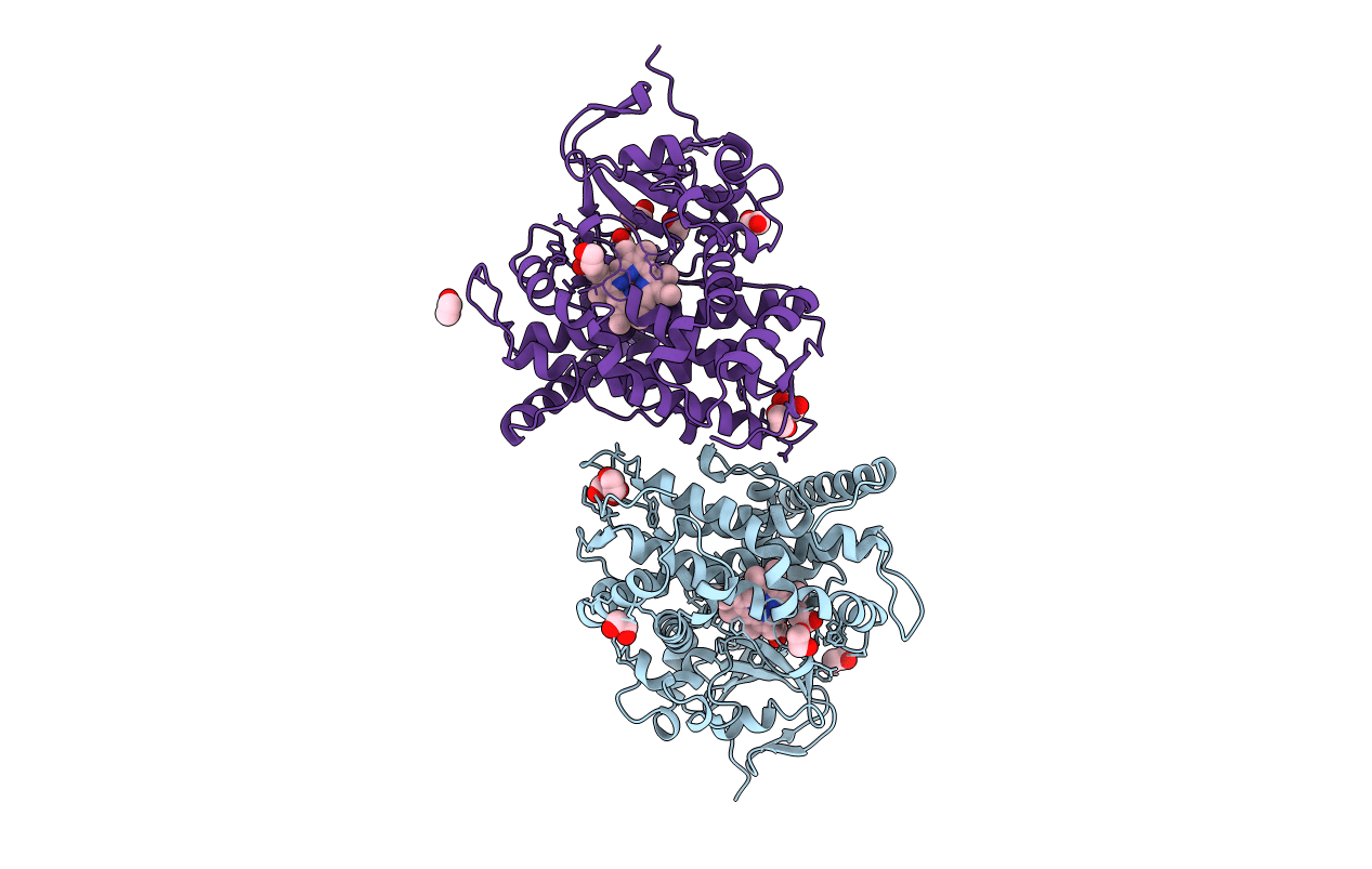
1BU7 image
Entry Detail
PDB ID:
1BU7
Keywords:
Title:
CRYOGENIC STRUCTURE OF CYTOCHROME P450BM-3 HEME DOMAIN
Biological Source:
Source Organism:
Host Organism:
PDB Version:
Deposition Date:
1998-09-14
Release Date:
1998-09-23
Method Details:
Experimental Method:
Resolution:
1.65 Å
R-Value Free:
0.25
R-Value Observed:
0.19
Space Group:
P 1 21 1
Macromolecular Entities
Polymer Type:polypeptide(L)
Description:PROTEIN (CYTOCHROME P450)
Chain IDs:A, B
Chain Length:455
Number of Molecules:2
Biological Source:Bacillus megaterium
Primary Citation
Structure of a cytochrome P450-redox partner electron-transfer complex.
Proc.Natl.Acad.Sci.USA 96 1863 1868 (1999)
PMID: 10051560 DOI: 10.1073/pnas.96.5.1863

Abstact

The crystal structure of the complex between the heme- and FMN-binding domains of bacterial cytochrome P450BM-3, a prototype for the complex between eukaryotic microsomal P450s and P450 reductase, has been determined at 2.03 A resolution. The flavodoxin-like flavin domain is positioned at the proximal face of the heme domain with the FMN 4.0 and 18.4 A from the peptide that precedes the heme-binding loop and the heme iron, respectively. The heme-binding peptide represents the most efficient and coupled through-bond electron pathway to the heme iron. Substantial differences between the FMN-binding domains of P450BM-3 and microsomal P450 reductase, observed around the flavin-binding sites, are responsible for different redox properties of the FMN, which, in turn, control electron flow to the P450.

Legend

Protein

Chemical

Disease

Primary Citation of related structures