1BU6 image
Deposition Date 1998-08-30
Release Date 1998-09-16
Last Version Date 2023-08-09
Entry Detail
PDB ID:
1BU6
Keywords:
Title:
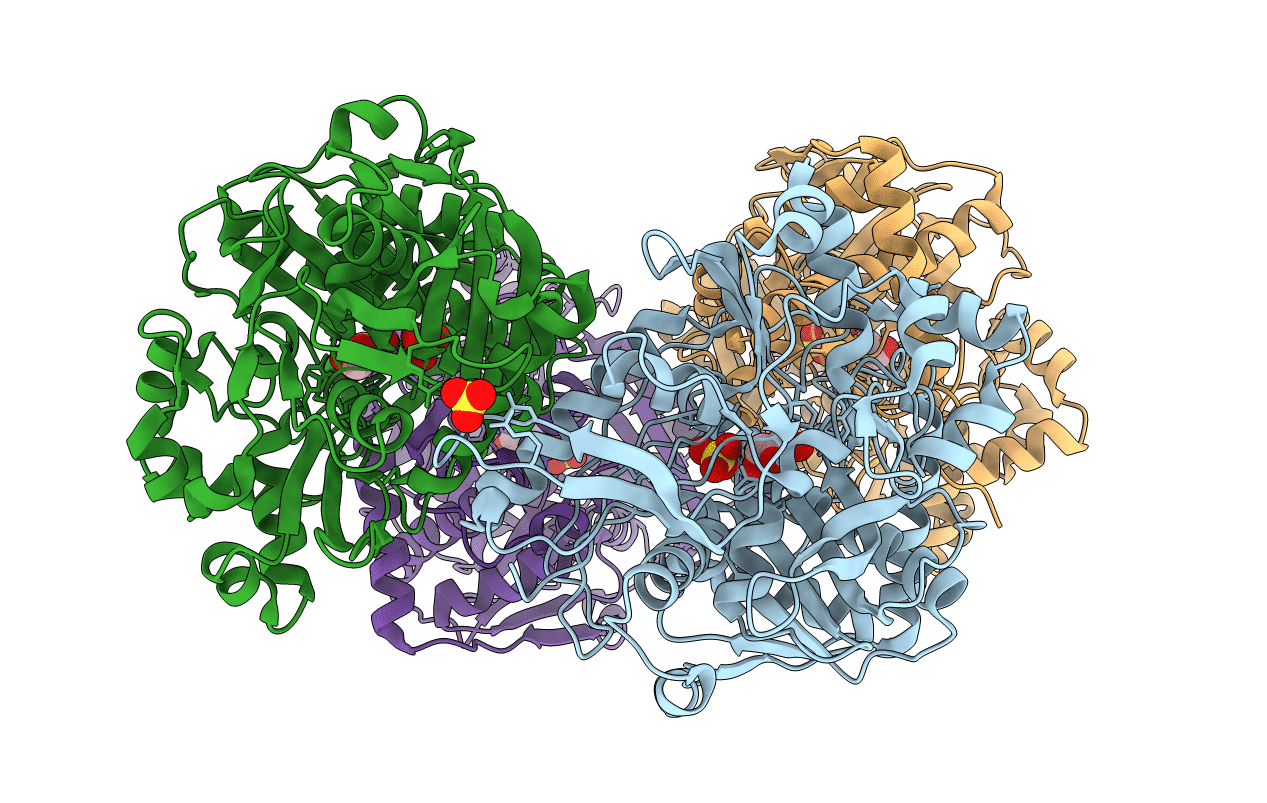
CRYSTAL STRUCTURES OF ESCHERICHIA COLI GLYCEROL KINASE AND THE MUTANT A65T IN AN INACTIVE TETRAMER: CONFORMATIONAL CHANGES AND IMPLICATIONS FOR ALLOSTERIC REGULATION
Biological Source:
Source Organism:
Escherichia coli (Taxon ID: 562)
Host Organism:
Method Details:
Experimental Method:
Resolution:
2.37 Å
R-Value Work:
0.16
Space Group:
P 1 21 1
Macromolecular Entities
Structures with similar UniProt ID
Protein Blast
Polymer Type:polypeptide(L)
Molecule:PROTEIN (GLYCEROL KINASE)
Gene (Uniprot):glpK
Mutagens:A65T
Chain IDs:A (auth: O), B (auth: Y), C (auth: Z), D (auth: X)
Chain Length:501
Number of Molecules:4
Biological Source:Escherichia coli
Primary Citation
Glycerol kinase from Escherichia coli and an Ala65-->Thr mutant: the crystal structures reveal conformational changes with implications for allosteric regulation.
Structure 6 1407 1418 (1998)
PMID: 9817843 DOI: 10.1016/S0969-2126(98)00140-3

Abstact

BACKGROUND Glycerol kinase (GK) from Escherichia coli is a velocity-modulated (V system) enzyme that has three allosteric effectors with independent mechanisms: fructose-1,6-bisphosphate (FBP); the phosphocarrier protein IIAGlc; and adenosine nucleotides. The enzyme exists in solution as functional dimers that associate reversibly to form tetramers. GK is a member of a superfamily of ATPases that share a common ATPase domain and are thought to undergo a large conformational change as an intrinsic step in their catalytic cycle. Members of this family include actin, hexokinase and the heat shock protein hsc70. RESULTS We report here the crystal structures of GK and a mutant of GK (Ala65-->Thr) in complex with glycerol and ADP. Crystals of both enzymes contain the same 222 symmetric tetramer. The functional dimer is identical to that described previously for the IIAGlc-GK complex structure. The tetramer interface is significantly different, however, with a relative 22.3 degrees rotation and 6.34 A translation of one functional dimer. The overall monomer structure is unchanged except for two regions: the IIAGlc-binding site undergoes a structural rearrangement and residues 230-236 become ordered and bind orthophosphate at the tetramer interface. We also report the structure of a second mutant of GK (IIe474-->Asp) in complex with IIAGlc; this complex crystallized isomorphously to the wild type IIAGlc-GK complex. Site-directed mutants of GK with substitutions at the IIAGlc-binding site show significantly altered kinetic and regulatory properties, suggesting that the conformation of the binding site is linked to the regulation of activity. CONCLUSIONS We conclude that the new tetramer structure presented here is an inactive form of the physiologically relevant tetramer. The structure and location of the orthophosphate-binding site is consistent with it being part of the FBP-binding site. Mutational analysis and the structure of the IIAGlc-GK(IIe474-->Asp) complex suggest the conformational transition of the IIAGlc-binding site to be an essential aspect of IIAGlc regulation.

Legend

Protein

Chemical

Disease

Primary Citation of related structures