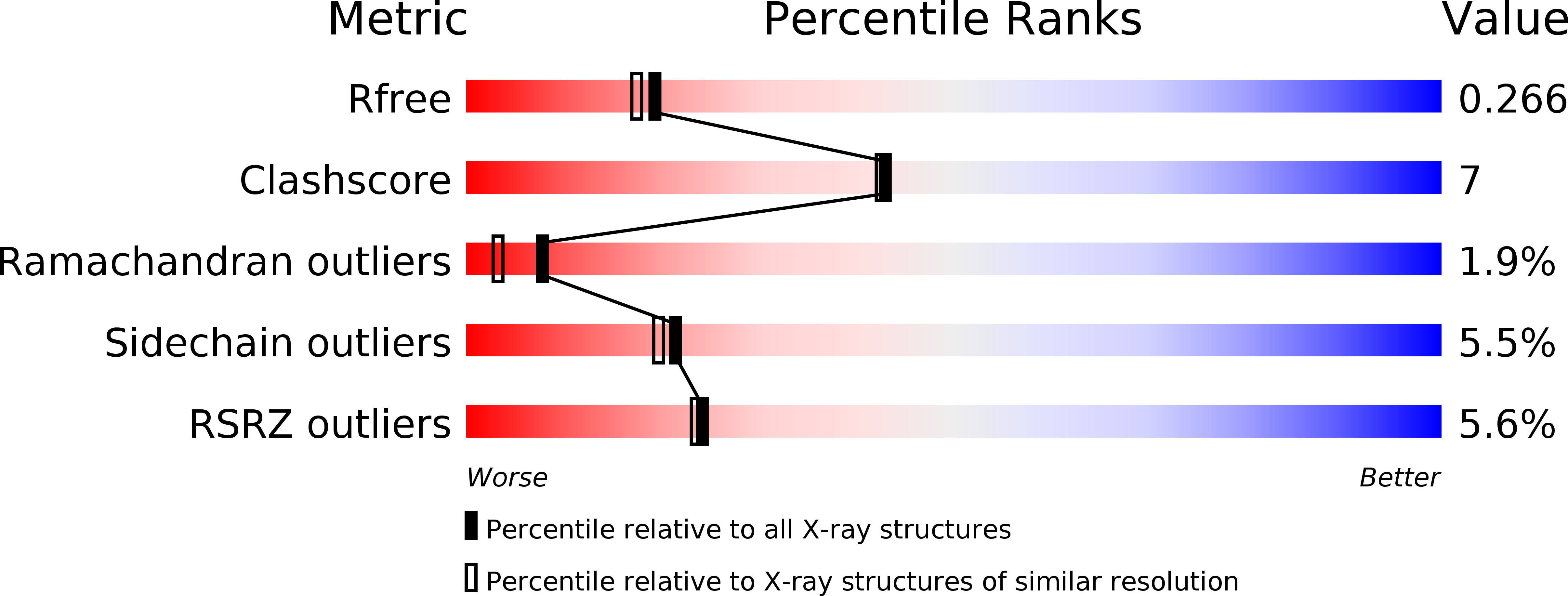
1BSO image
Deposition Date 1998-08-29
Release Date 1999-09-02
Last Version Date 2024-10-30
Entry Detail
PDB ID:
1BSO
Title:
12-BROMODODECANOIC ACID BINDS INSIDE THE CALYX OF BOVINE BETA-LACTOGLOBULIN
Biological Source:
Source Organism:
Bos taurus (Taxon ID: 9913)
Method Details:
Experimental Method:
Resolution:
2.23 Å
R-Value Free:
0.27
R-Value Work:
0.23
R-Value Observed:
0.23
Space Group:
P 32 2 1
Macromolecular Entities
Polymer Type:polypeptide(L)
Molecule:PROTEIN (BOVINE BETA-LACTOGLOBULIN A)
Gene (Uniprot):LGB
Chain IDs:A
Chain Length:162
Number of Molecules:1
Biological Source:Bos taurus
Ligand Molecules
Primary Citation
12-Bromododecanoic acid binds inside the calyx of bovine beta-lactoglobulin.
FEBS Lett. 438 272 278 (1998)
PMID: 9827560 DOI: 10.1016/S0014-5793(98)01199-5

Abstact

The X-ray structure of bovine beta-lactoglobulin with the ligand 12-bromododecanoic acid as a model for fatty acids has been determined at a resolution of 2.23 A in the trigonal lattice Z form. The ligand binds inside the calyx, resolving a long-standing controversy as to where fatty-acid like ligands bind. The carboxylate head group lies at the surface of the molecule, and the lid to the calyx is open at the pH of crystallization (pH 7.3), consistent with the conformation observed in ligand-free bovine beta-lactoglobulin in lattice Z at pH 7.1 and pH 8.2.

Legend

Protein

Chemical

Disease

Primary Citation of related structures