1BS4 image
Entry Detail
PDB ID:
1BS4
Keywords:
Title:
PEPTIDE DEFORMYLASE AS ZN2+ CONTAINING FORM (NATIVE) IN COMPLEX WITH INHIBITOR POLYETHYLENE GLYCOL
Biological Source:
Source Organism:
Host Organism:
PDB Version:
Deposition Date:
1998-09-01
Release Date:
1999-08-27
Method Details:
Experimental Method:
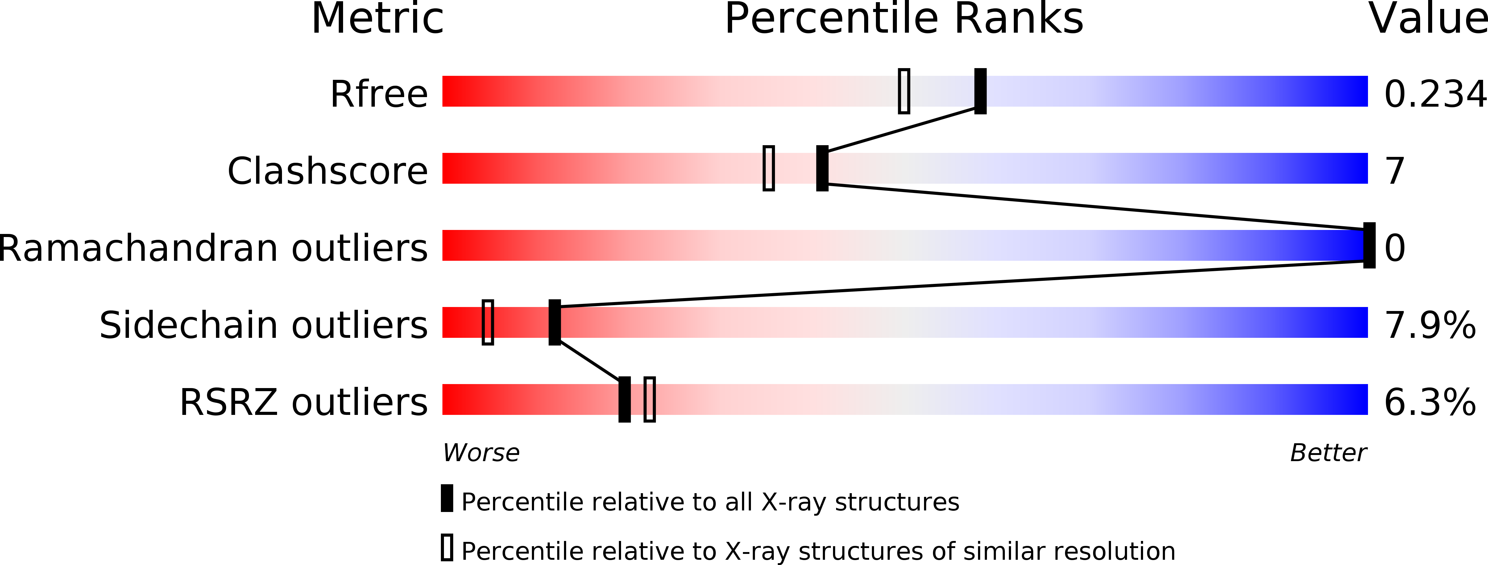
Resolution:
1.90 Å
R-Value Free:
0.24
R-Value Work:
0.19
R-Value Observed:
0.19
Space Group:
C 1 2 1
Macromolecular Entities
Polymer Type:polypeptide(L)
Description:PROTEIN (PEPTIDE DEFORMYLASE)
Chain IDs:A, B, C
Chain Length:168
Number of Molecules:3
Biological Source:Escherichia coli
Primary Citation
Iron center, substrate recognition and mechanism of peptide deformylase.
Nat.Struct.Biol. 5 1053 1058 (1998)
PMID: 9846875 DOI: 10.1038/4162

Abstact

Eubacterial proteins are synthesized with a formyl group at the N-terminus which is hydrolytically removed from the nascent chain by the mononuclear iron enzyme peptide deformylase. Catalytic efficiency strongly depends on the identity of the bound metal. We have determined by X-ray crystallography the Fe2+, Ni2+ and Zn2+ forms of the Escherichia coli enzyme and a structure in complex with the reaction product Met-Ala-Ser. The structure of the complex, with the tripeptide bound at the active site, suggests detailed models for the mechanism of substrate recognition and catalysis. Differences of the protein structures due to the identity of the bound metal are extremely small and account only for the observation that Zn2+ binds more tightly than Fe2+ or Ni2+. The striking loss of catalytic activity of the Zn2+ form could be caused by its reluctance to change between tetrahedral and five-fold metal coordination believed to occur during catalysis. N-terminal formylation and subsequent deformylation

Legend

Protein

Chemical

Disease

Primary Citation of related structures