Deposition Date
1998-08-25
Release Date
1999-01-13
Last Version Date
2023-08-09
Entry Detail
PDB ID:
1BRW
Keywords:
Title:
THE CRYSTAL STRUCTURE OF PYRIMIDINE NUCLEOSIDE PHOSPHORYLASE IN A CLOSED CONFORMATION
Biological Source:
Source Organism(s):
Geobacillus stearothermophilus (Taxon ID: 1422)
Method Details:
Experimental Method:
Resolution:
2.10 Å
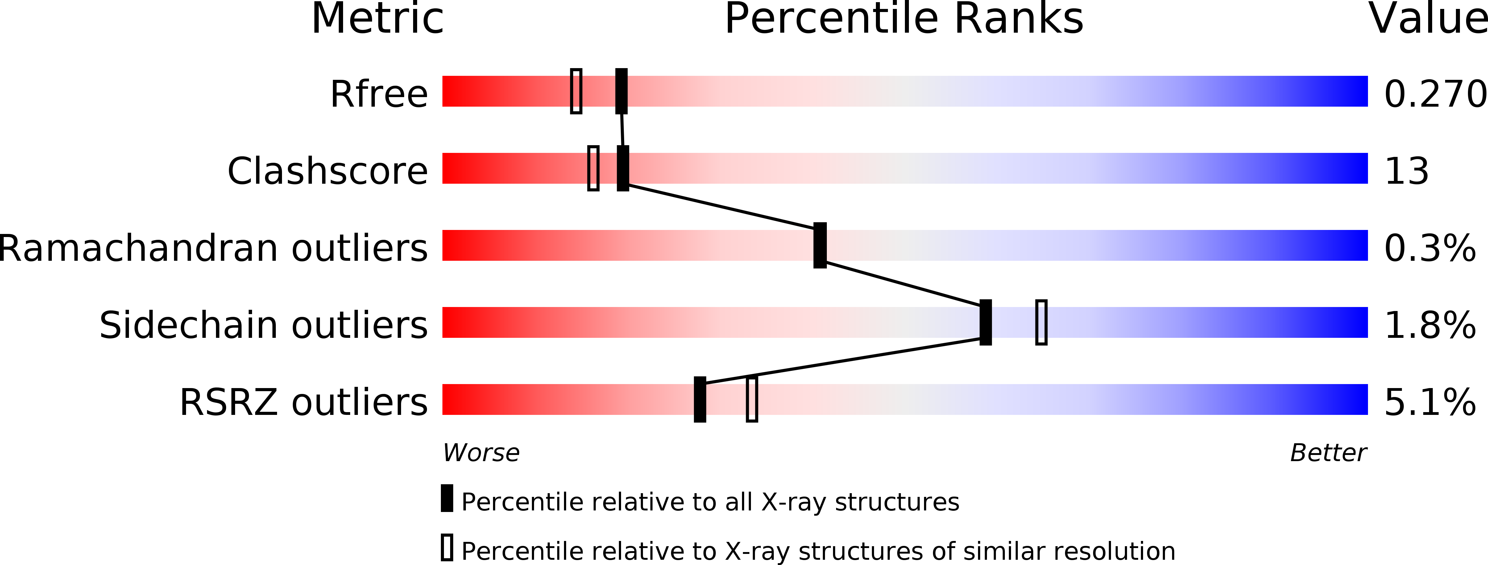
R-Value Free:
0.27
R-Value Work:
0.23
R-Value Observed:
0.23
Space Group:
P 1 21 1