1BRR image
Deposition Date 1998-07-28
Release Date 1998-09-30
Last Version Date 2024-11-20
Entry Detail
PDB ID:
1BRR
Title:
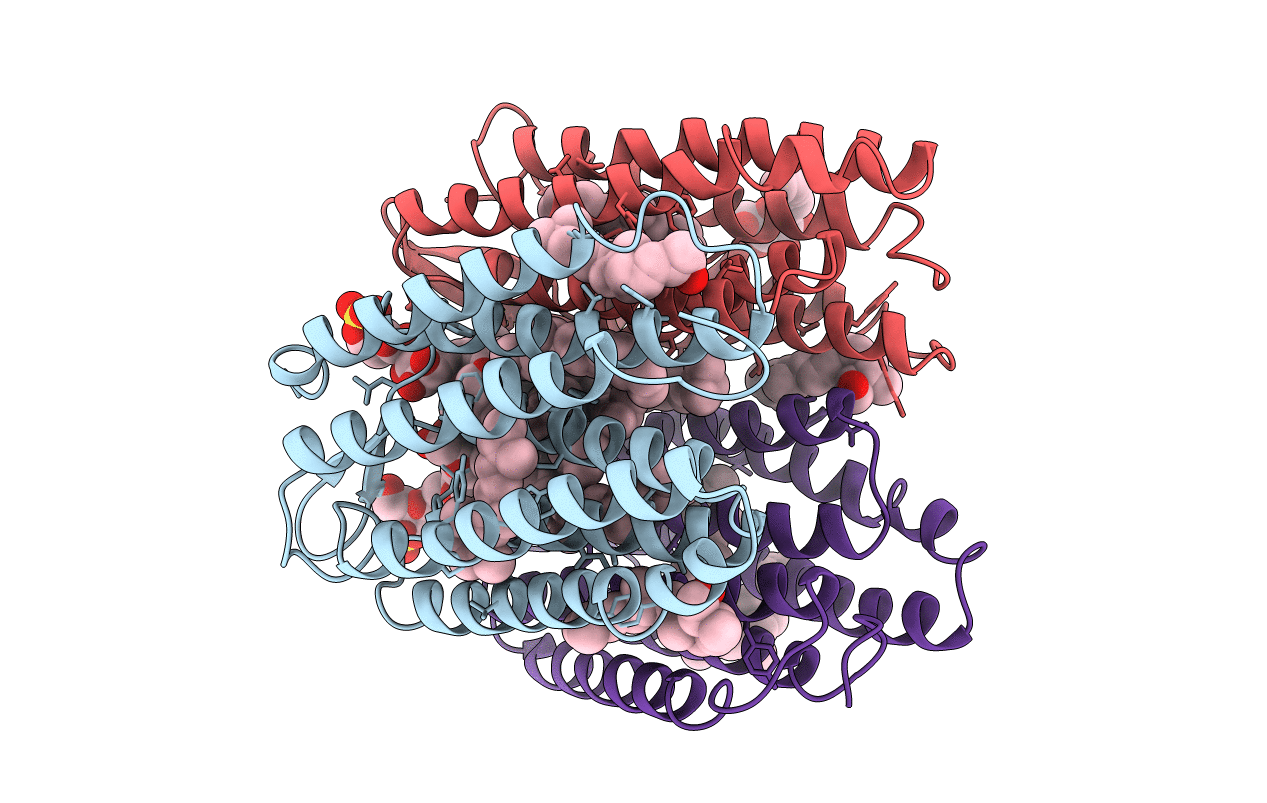
X-RAY STRUCTURE OF THE BACTERIORHODOPSIN TRIMER/LIPID COMPLEX
Biological Source:
Source Organism:
Method Details:
Experimental Method:
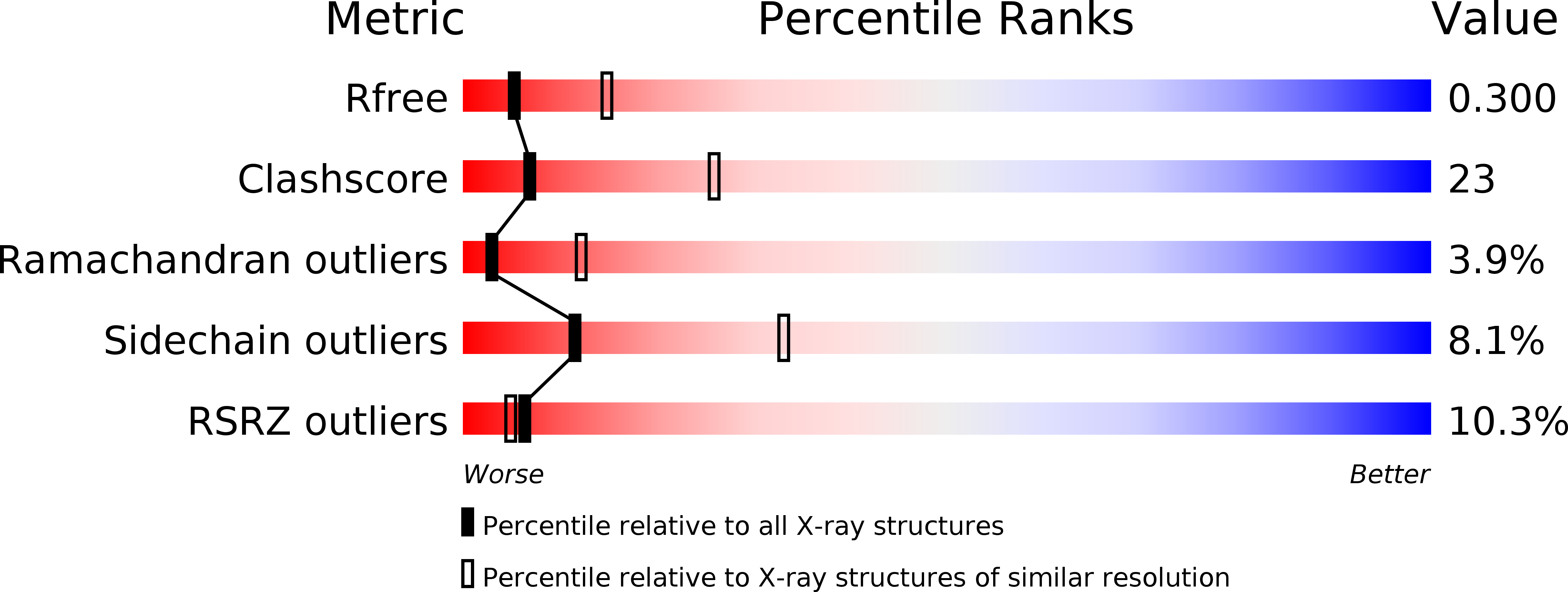
Resolution:
2.90 Å
R-Value Free:
0.29
R-Value Work:
0.25
R-Value Observed:
0.25
Space Group:
C 1 2 1
Macromolecular Entities
Polymer Type:polypeptide(L)
Molecule:PROTEIN (BACTERIORHODOPSIN)
Gene (Uniprot):bop
Chain IDs:A, B, C
Chain Length:247
Number of Molecules:3
Biological Source:Halobacterium salinarum
Modified Residue
Compound ID Chain ID Parent Comp ID Details 2D Image
PCA C GLN PYROGLUTAMIC ACID
Primary Citation
Lipid patches in membrane protein oligomers: crystal structure of the bacteriorhodopsin-lipid complex
Proc.Natl.Acad.Sci.USA 95 11673 11678 (1998)
PMID: 9751724 DOI: 10.1073/pnas.95.20.11673

Abstact

Heterogenous nucleation on small molecule crystals causes a monoclinic crystal form of bacteriorhodopsin (BR) in which trimers of this membrane protein pack differently than in native purple membranes. Analysis of single crystals by nano-electrospray ionization-mass spectrometry demonstrated a preservation of the purple membrane lipid composition in these BR crystals. The 2.9-A x-ray structure shows a lipid-mediated stabilization of BR trimers where the glycolipid S-TGA-1 binds into the central compartment of BR trimers. The BR trimer/lipid complex provides an example of local membrane thinning as the lipid head-group boundary of the central lipid patch is shifted by 5 A toward the membrane center. Nonbiased electron density maps reveal structural differences to previously reported BR structures, especially for the cytosolic EF loop and the proton exit pathway. The terminal proton release complex now comprises an E194-E204 dyad as a diffuse proton buffer.

Legend

Protein

Chemical

Disease

Primary Citation of related structures