Deposition Date
1998-08-25
Release Date
1998-09-02
Last Version Date
2024-05-22
Entry Detail
PDB ID:
1BR0
Keywords:
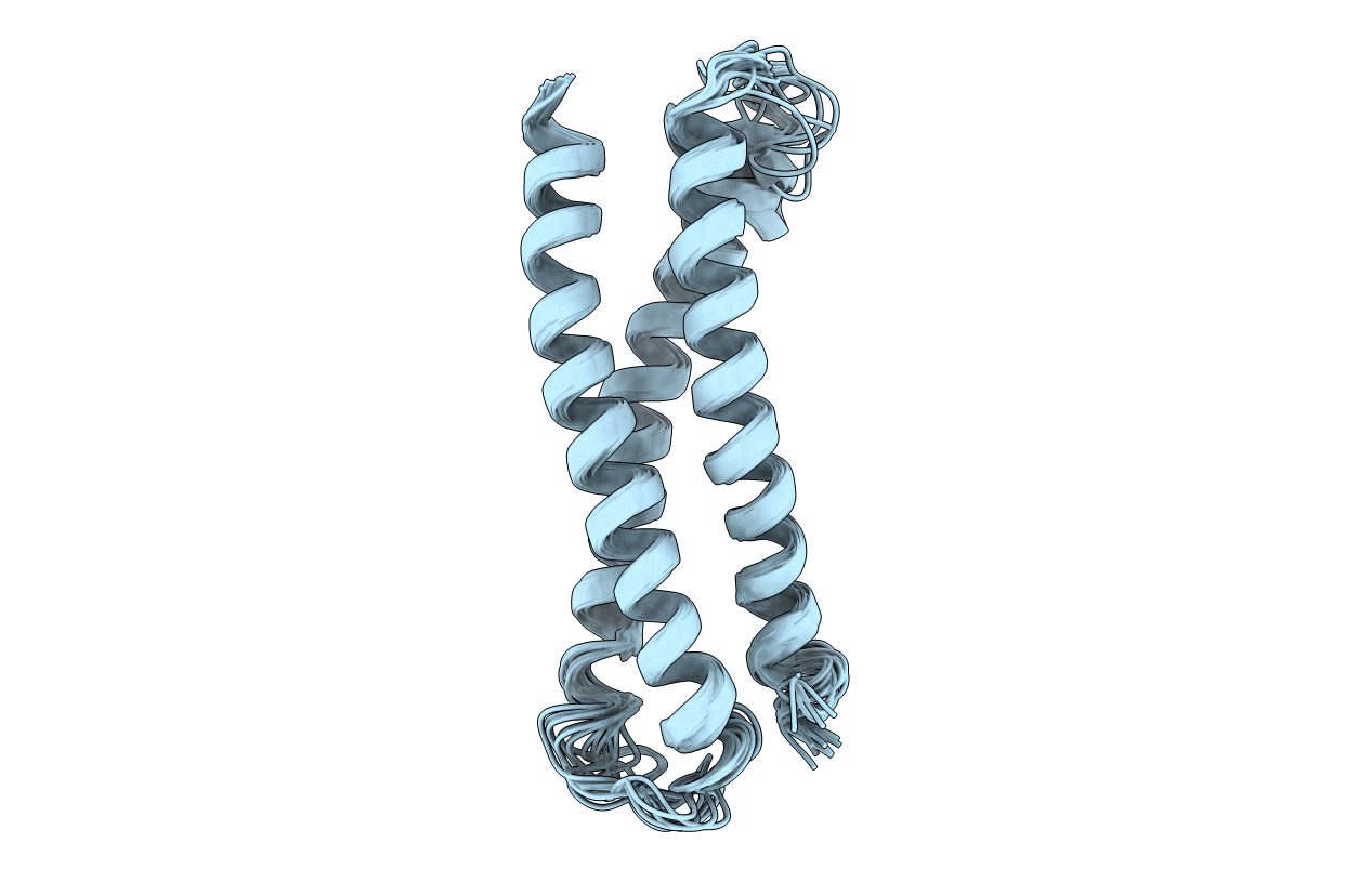
Title:
THREE DIMENSIONAL STRUCTURE OF THE N-TERMINAL DOMAIN OF SYNTAXIN 1A
Biological Source:
Source Organism(s):
Rattus norvegicus (Taxon ID: 10116)
Expression System(s):
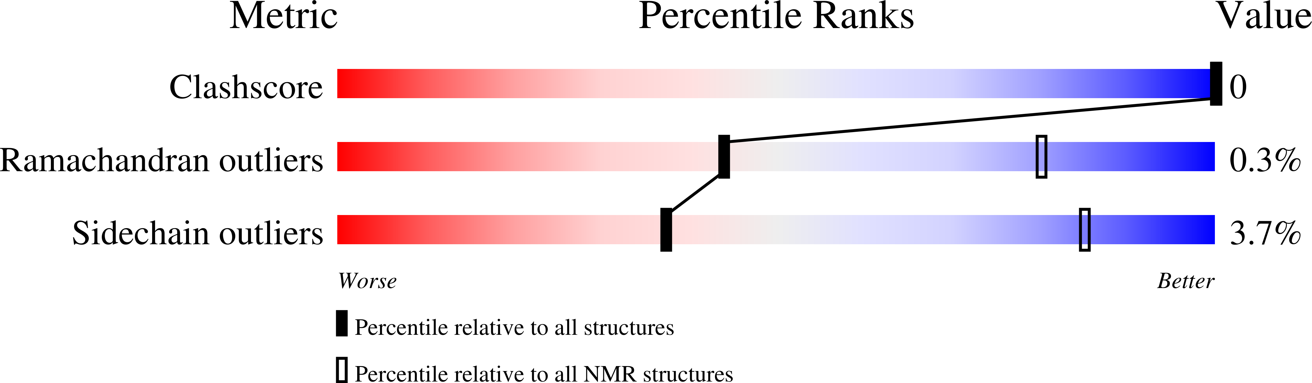
Method Details:
Experimental Method:
Conformers Calculated:
200
Conformers Submitted:
15
Selection Criteria:
LEAST RESTRAINT VIOLATION