1BQS image
Deposition Date 1998-08-18
Release Date 1999-08-13
Last Version Date 2024-11-13
Entry Detail
PDB ID:
1BQS
Title:
THE CRYSTAL STRUCTURE OF MUCOSAL ADDRESSIN CELL ADHESION MOLECULE-1 (MADCAM-1)
Biological Source:
Source Organism:
Homo sapiens (Taxon ID: 9606)
Method Details:
Experimental Method:
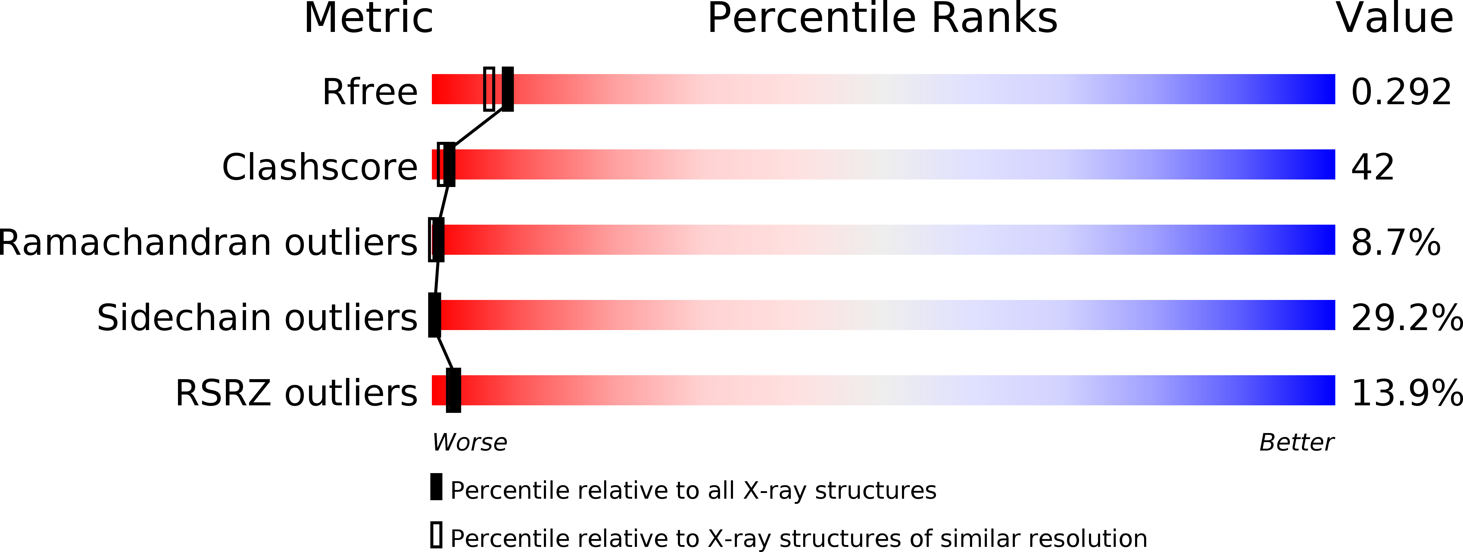
Resolution:
2.20 Å
R-Value Free:
0.28
R-Value Work:
0.22
R-Value Observed:
0.22
Space Group:
C 2 2 21
Macromolecular Entities
Polymer Type:polypeptide(L)
Molecule:PROTEIN (MUCOSAL ADDRESSIN CELL ADHESION MOLECULE-1)
Gene (Uniprot):MADCAM1
Chain IDs:A
Chain Length:209
Number of Molecules:1
Biological Source:Homo sapiens
Modified Residue
Compound ID Chain ID Parent Comp ID Details 2D Image
ASN A ASN GLYCOSYLATION SITE
Ligand Molecules
Primary Citation
The structure of immunoglobulin superfamily domains 1 and 2 of MAdCAM-1 reveals novel features important for integrin recognition.
Structure 6 793 801 (1998)
PMID: 9655832 DOI: 10.1016/S0969-2126(98)00080-X

Abstact

BACKGROUND Mucosal addressin cell adhesion molecule 1 (MAdCAM-1) is a cell adhesion molecule that is expressed on the endothelium in mucosa, and guides the specific homing of lymphocytes into mucosal tissues. MAdCAM-1 belongs to a subclass of the immunoglobulin superfamily (IgSF), the members of which are ligands for integrins. Human MAdCAM-1 has a unique dual function compared to other members in the same subclass in that it binds both the integrin alpha4beta7, through its two IgSF domains, and a selectin expressed on leukocytes, via carbohydrate sidechains. The structure determination of the two IgSF domains and comparison to the N-terminal two-domain structures of vascular cell adhesion molecule 1 (VCAM-1) and intercellular adhesion molecules (ICAM-1 and ICAM-2) allow us to assess the molecular basis of the interactions between integrins and their preferred ligands. RESULTS The crystal structure of a fragment containing the two IgSF domains of human MAdCAM-1 has been determined to 2.2 A resolution. The structure of MAdCAM-1 reveals two separate integrin-recognition motifs. The key integrin-binding residue, Asp42, resides in the CD loop of domain 1; a buried arginine residue (Arg70) plays a critical role in maintaining the conformation of this loop. The second binding site is associated with an unusual long D strand in domain 2. The D and E strands extend beyond the main body of the domain, forming a negatively charged beta ribbon unique to MAdCAM-1. This ribbon is located on the same face as the key aspartate residue in domain 1, consistent with evidence that it is involved in integrin binding. CONCLUSIONS The structural comparison of MAdCAM-1 to other members of the same IgSF subclass reveals some interesting features. Firstly, MAdCAM-1, like VCAM-1, has the key integrin-binding residue located on the protruding CD loop of domain 1 and binds to an integrin that lacks an I domain. This is in contrast to ICAM-1 and ICAM-2 where the key residue is located at the end of the C strand on a flat surface and which bind to integrins that contain I domains. Secondly, architectural differences in the CD loops of MAdCAM-1 and VCAM-1 cause an 8 A shift in position of the critical aspartate residue, and may partly determine their binding preference for different integrins. Finally, the unusual charge distribution of the two-domain fragment of MAdCAM-1 is predicted to orient the molecule optimally for integrin binding on the top of its long mucin-like stalk.

Legend

Protein

Chemical

Disease

Primary Citation of related structures