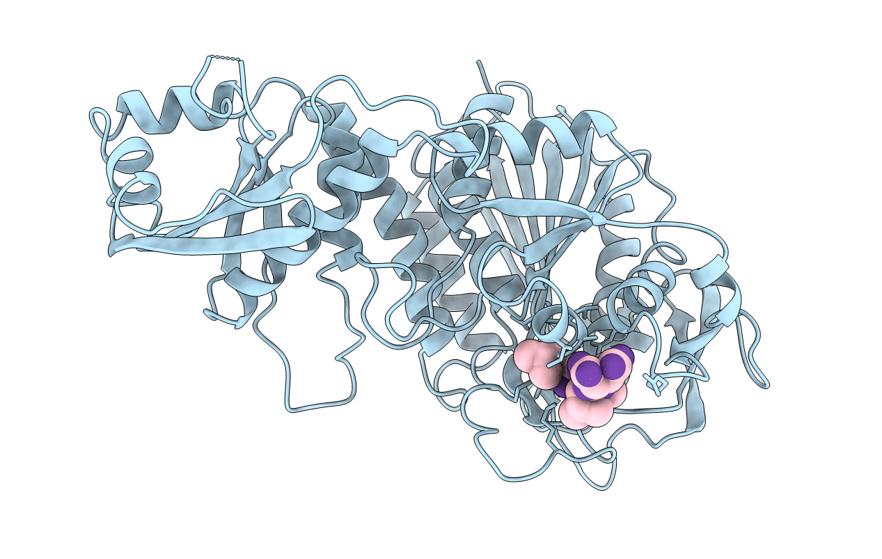
1BLL image
Deposition Date 1993-03-02
Release Date 1994-01-31
Last Version Date 2024-10-30
Entry Detail
PDB ID:
1BLL
Title:
X-RAY CRYSTALLOGRAPHIC DETERMINATION OF THE STRUCTURE OF BOVINE LENS LEUCINE AMINOPEPTIDASE COMPLEXED WITH AMASTATIN: FORMULATION OF A CATALYTIC MECHANISM FEATURING A GEM-DIOLATE TRANSITION STATE
Biological Source:
Source Organism:
Bos taurus (Taxon ID: 9913)
Streptomyces sp. ME98-M3 (Taxon ID: )
Method Details:
Experimental Method:
Resolution:
2.40 Å
R-Value Work:
0.19
R-Value Observed:
0.19
Space Group:
P 63 2 2
Macromolecular Entities
Structures with similar UniProt ID
Protein Blast
Polymer Type:polypeptide(L)
Molecule:LEUCINE AMINOPEPTIDASE
Gene (Uniprot):LAP3
Chain IDs:A (auth: E)
Chain Length:488
Number of Molecules:1
Biological Source:Bos taurus
Polymer Type:polypeptide(L)
Molecule:AMASTATIN
Chain IDs:B (auth: I)
Chain Length:4
Number of Molecules:1
Biological Source:Streptomyces sp. ME98-M3
Ligand Molecules
Peptide-like Molecules
PRD_000415
Primary Citation
X-ray crystallographic determination of the structure of bovine lens leucine aminopeptidase complexed with amastatin: formulation of a catalytic mechanism featuring a gem-diolate transition state.
Biochemistry 32 8465 8478 (1993)
PMID: 8357796 DOI: 10.1021/bi00084a011

Abstact

The structure of the complex of bovine lens leucine aminopeptidase (blLAP) with the slow-, tight-binding inhibitor amastatin has been determined by X-ray crystallography. X-ray diffraction data were collected at -150 degrees C from a single blLAP-amastatin crystal which under the data collection conditions was of the space group P6(3)22 with unit cell parameters a = 130.3 A and c = 121.9 A. The structure of the blLAP-amastatin complex was determined by molecular replacement, using the structure of native blLAP as the starting model. Refinement of the blLAP-amastatin model plus 132 water molecules against data from 10.0- to 2.4-A resolution resulted in a final structure with a crystallographic residual of 0.198. The binding mode of amastatin is similar to that of bestatin, the structure of whose complex with blLAP has previously been determined. Of particular note, the N-terminus-to-C-terminus orientation of the two bound inhibitors is the same. The two N-terminal residues of amastatin and bestatin occupy the same binding sites, which are most likely S1 and S'1. The slow binding of amastatin and bestatin may be partially attributable to a binding mechanism in which the two active site metals are sequentially coordinated by the P1 amino and hydroxyl groups of these inhibitors. A catalytic mechanism for blLAP is proposed based on the binding modes of amastatin and bestatin and plausible binding modes of a dipeptide substrate and its putative gem-diolate transition state which were modeled into the active site of blLAP after the binding mode of amastatin. The proposed catalytic mechanism invokes roles for the catalytic metals in binding and activating the substrate and in stabilizing the transition state. The mechanism also includes roles for Asp-255 as a general base, Arg-336 as an additional electrophilic substrate activator and transition state stabilizer, and Lys-262 as a proton shuttle.

Legend

Protein

Chemical

Disease

Primary Citation of related structures