1BLF image
Deposition Date 1997-08-20
Release Date 1997-12-03
Last Version Date 2024-10-23
Entry Detail
PDB ID:
1BLF
Title:
STRUCTURE OF DIFERRIC BOVINE LACTOFERRIN AT 2.8 ANGSTROMS RESOLUTION
Biological Source:
Source Organism:
Bos taurus (Taxon ID: 9913)
Method Details:
Experimental Method:
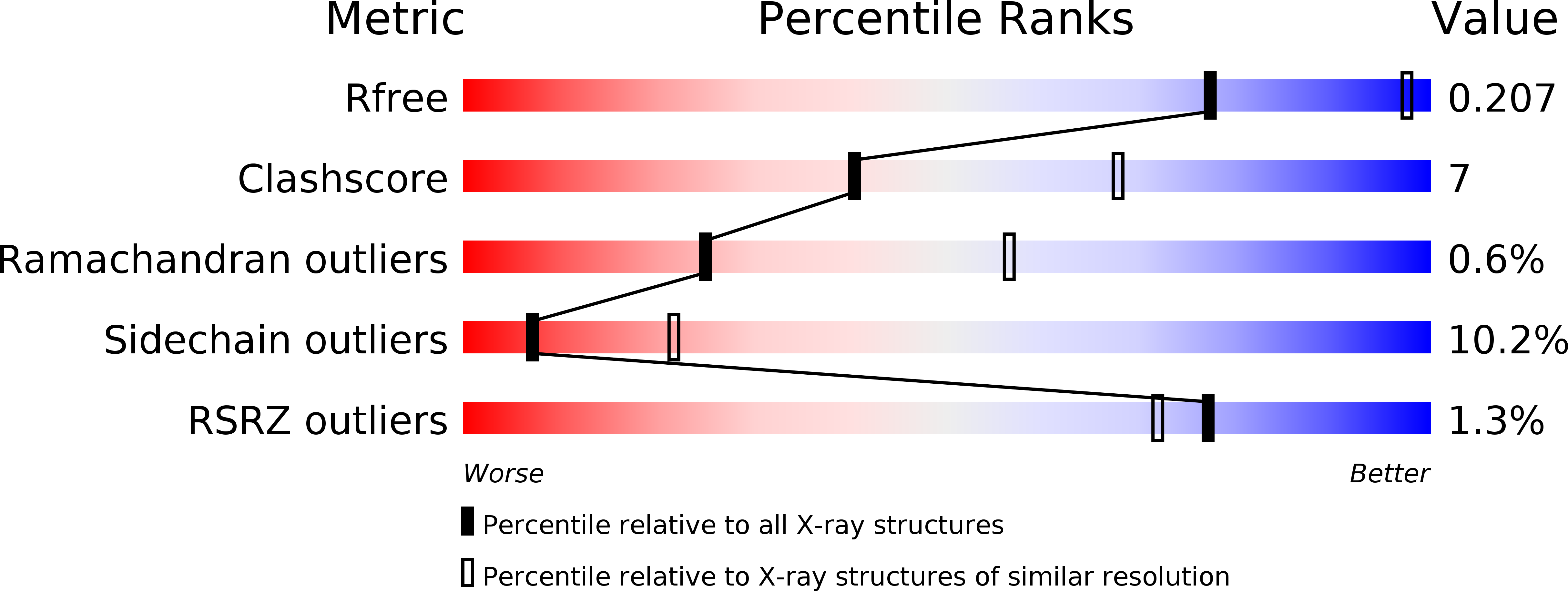
Resolution:
2.80 Å
R-Value Free:
0.28
R-Value Work:
0.21
R-Value Observed:
0.23
Space Group:
P 21 21 21
Macromolecular Entities
Polymer Type:polypeptide(L)
Molecule:LACTOFERRIN
Gene (Uniprot):LTF
Chain IDs:A
Chain Length:689
Number of Molecules:1
Biological Source:Bos taurus
Modified Residue
Compound ID Chain ID Parent Comp ID Details 2D Image
ASN A ASN GLYCOSYLATION SITE
Primary Citation
Three-dimensional structure of diferric bovine lactoferrin at 2.8 A resolution.
J.Mol.Biol. 274 222 236 (1997)
PMID: 9398529 DOI: 10.1006/jmbi.1997.1386

Abstact

The three-dimensional structure of diferric bovine lactoferrin (bLf) has been determined by X-ray crystallography in order to investigate the factors that influence iron binding and release by transferrins. The structure was solved by molecular replacement, using the coordinates of diferric human lactoferrin (hLf) as a search model, and was refined with data to 2.8 A resolution by simulated annealing (X-PLOR) and restrained least squares (TNT). The final model comprises 5310 protein atoms (residues 5 to 689), 124 carbohydrate atoms (from ten monosaccharide units, in three glycan chains), 2 Fe3+, 2 CO32- and 50 water molecules. This model gives an R-factor of 0.232 for 21440 reflections in the resolution range 30.0 to 2.8 A. The folding of the bLf molecule is essentially the same as that of hLf, but bLf differs in the extent of closure of the two domains of each lobe, and in the relative orientations of the two lobes. Differences in domain closure are attributed to amino acid changes in the interface, and differences in lobe orientations to slightly altered packing of two hydrophobic patches between the lobes. Changed interdomain interactions may explain the lesser iron affinity of bLf, compared with hLf, and two lysine residues behind the N-lobe iron site of bLf offer new insights into the "dilysine trigger" mechanism proposed for iron release by transferrins. The bLf structure is also notable for several well-defined oligosaccharide units which demonstrate the structural factors that stabilise carbohydrate structure. One glycan chain, attached to Asn545, appears to contribute to interdomain interactions and may modulate iron release from the C-lobe.

Legend

Protein

Chemical

Disease

Primary Citation of related structures