Deposition Date
1998-07-10
Release Date
1998-07-15
Last Version Date
2024-02-07
Entry Detail
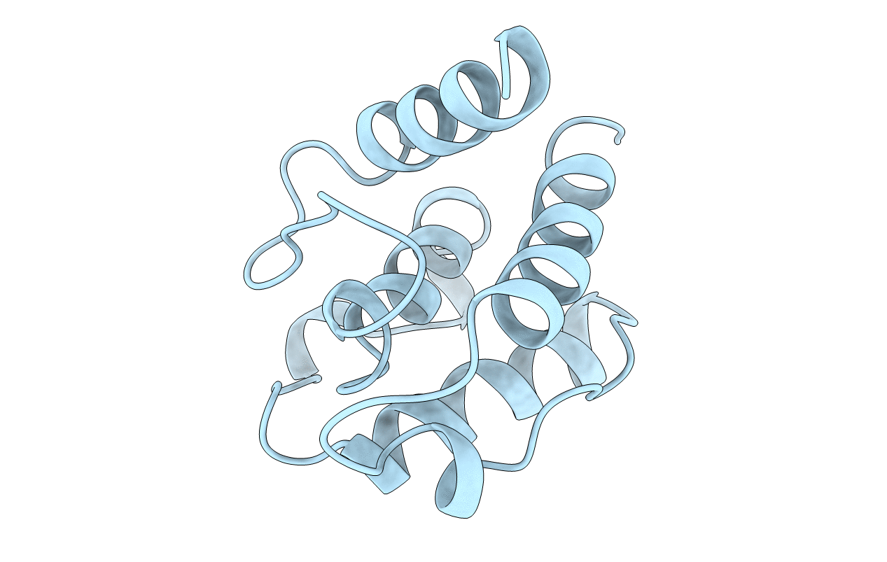
PDB ID:
1BKR
Keywords:
Title:
CALPONIN HOMOLOGY (CH) DOMAIN FROM HUMAN BETA-SPECTRIN AT 1.1 ANGSTROM RESOLUTION
Biological Source:
Source Organism:
Homo sapiens (Taxon ID: 9606)
Host Organism:
Method Details:
Experimental Method:
Resolution:
1.10 Å
R-Value Free:
0.18
R-Value Observed:
0.14
Space Group:
P 1 21 1