
Deposition Date
1995-09-27
Release Date
1996-01-29
Last Version Date
2024-10-16
Entry Detail
PDB ID:
1BIM
Keywords:
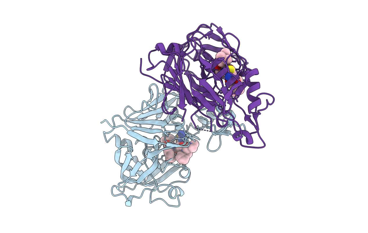
Title:
CRYSTALLOGRAPHIC STUDIES ON THE BINDING MODES OF P2-P3 BUTANEDIAMIDE RENIN INHIBITORS
Biological Source:
Source Organism(s):
Homo sapiens (Taxon ID: 9606)
Expression System(s):
Method Details:
Experimental Method:
Resolution:
2.80 Å
R-Value Work:
0.17
R-Value Observed:
0.17
Space Group:
P 21 3
