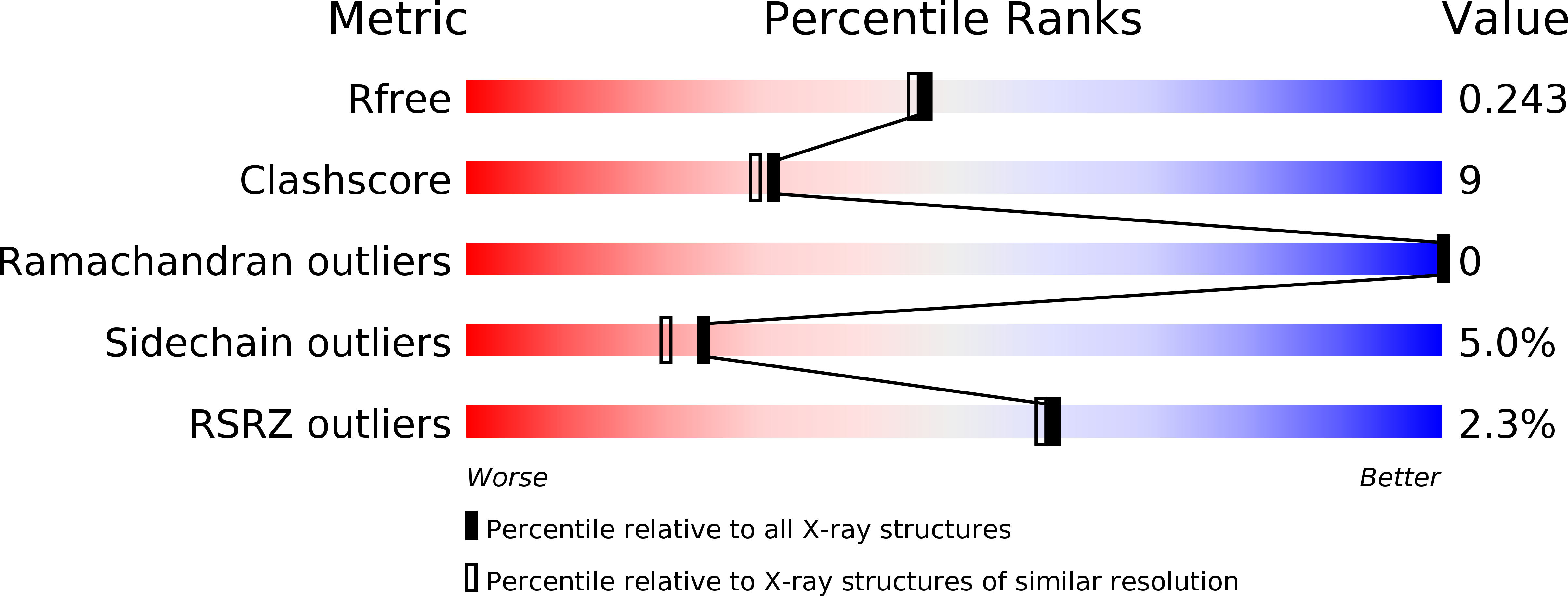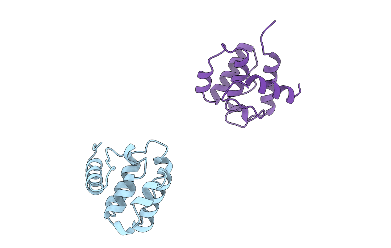
1BHD image
Deposition Date 1998-06-05
Release Date 1999-01-13
Last Version Date 2024-05-22
Entry Detail
PDB ID:
1BHD
Title:
SECOND CALPONIN HOMOLOGY DOMAIN FROM UTROPHIN
Biological Source:
Source Organism:
Homo sapiens (Taxon ID: 9606)
Method Details:
Experimental Method:
Resolution:
2.00 Å
R-Value Free:
0.25
R-Value Work:
0.18
R-Value Observed:
0.20
Space Group:
P 1 21 1
Macromolecular Entities
Structures with similar UniProt ID
Protein Blast
Polymer Type:polypeptide(L)
Molecule:UTROPHIN
Gene (Uniprot):UTRN
Chain IDs:A, B
Chain Length:118
Number of Molecules:2
Biological Source:Homo sapiens
Primary Citation
The 2.0 A structure of the second calponin homology domain from the actin-binding region of the dystrophin homologue utrophin.
J.Mol.Biol. 285 1257 1264 (1999)
PMID: 9887274 DOI: 10.1006/jmbi.1998.2406

Abstact

Utrophin is a close homologue of dystrophin, the protein defective in Duchenne muscular dystrophy. Like dystrophin, it is composed of three regions: an N-terminal region that binds actin filaments, a large central region with triple coiled-coil repeats, and a C-terminal region that interacts with components in the dystroglycan protein complex at the plasma membrane. The N-terminal actin-binding region consists of two calponin homology domains and is related to the actin-binding domains of a superfamily of proteins including alpha-actinin, spectrin and fimbrin. Here, we present the 2.0 A structure of the second calponin homology domain of utrophin solved by X-ray crystallography, and compare it to the other calponin homology domains previously determined from spectrin and fimbrin.

Legend

Protein

Chemical

Disease

Primary Citation of related structures