
Deposition Date
1994-06-09
Release Date
1994-09-30
Last Version Date
2024-02-07
Entry Detail
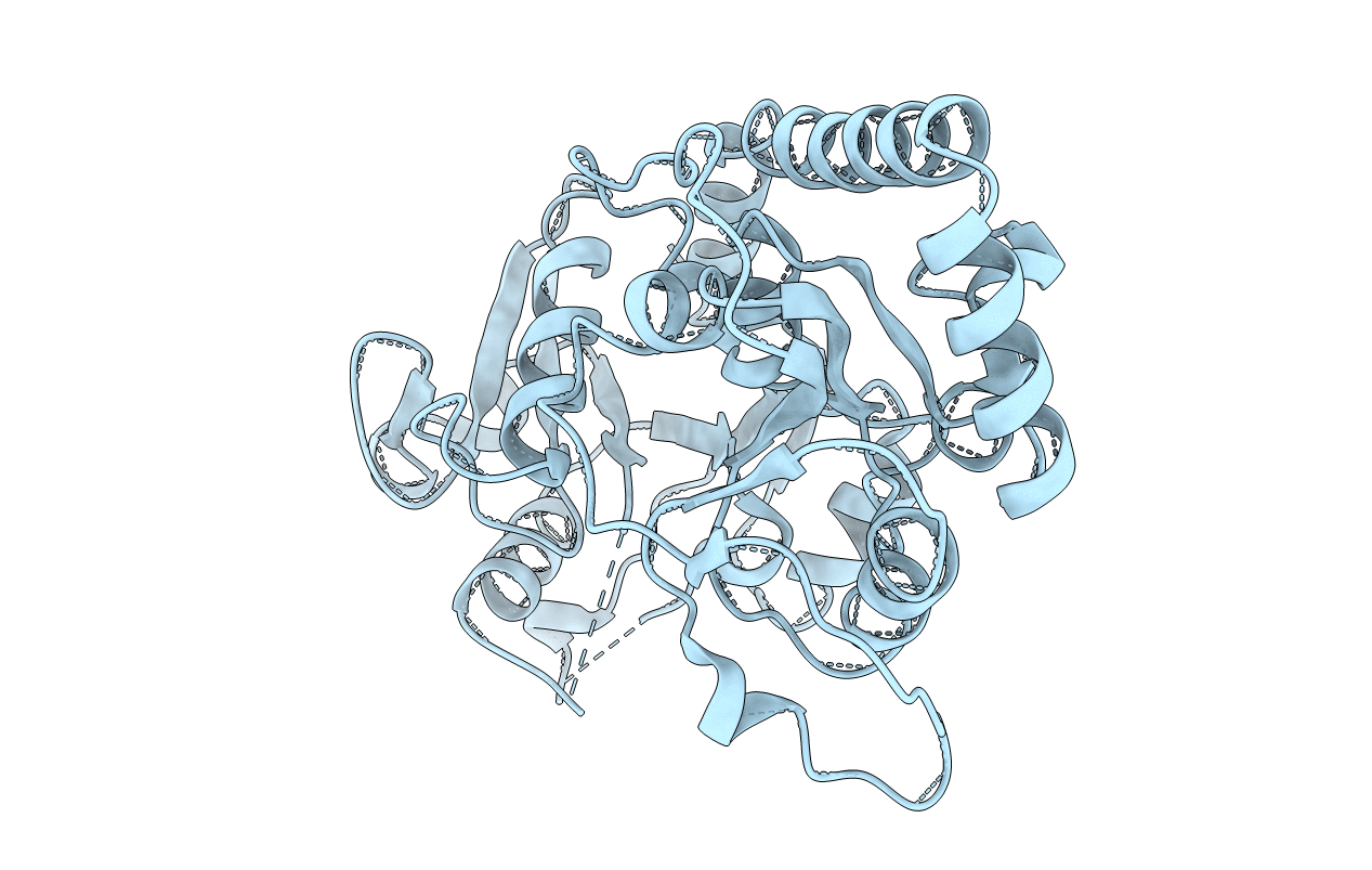
PDB ID:
1BGT
Keywords:
Title:
CRYSTAL STRUCTURE OF THE DNA MODIFYING ENZYME BETA-GLUCOSYLTRANSFERASE IN THE PRESENCE AND ABSENCE OF THE SUBSTRATE URIDINE DIPHOSPHOGLUCOSE
Biological Source:
Source Organism(s):
Enterobacteria phage T4 (Taxon ID: 10665)
Method Details:
Experimental Method:
Resolution:
2.20 Å
R-Value Work:
0.19
R-Value Observed:
0.19
Space Group:
P 21 21 2
