Deposition Date
1997-09-12
Release Date
1998-01-28
Last Version Date
2025-02-26
Entry Detail
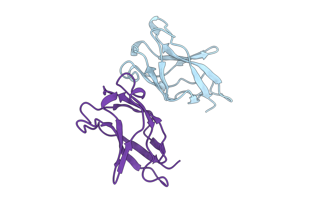
PDB ID:
1BFT
Keywords:
Title:
STRUCTURE OF NF-KB P65 HOMODIMER BOUND TO A KB SITE
Biological Source:
Source Organism(s):
Mus musculus (Taxon ID: 10090)
Method Details:
Experimental Method:
Resolution:
2.00 Å
R-Value Free:
0.29
R-Value Work:
0.20
R-Value Observed:
0.20
Space Group:
C 2 2 21