Deposition Date
1998-05-21
Release Date
1998-10-28
Last Version Date
2024-05-22
Entry Detail
PDB ID:
1BFK
Keywords:
Title:
CRYSTAL STRUCTURE OF SUBTILISIN CARLSBERG IN 40% ACETONITRILE
Biological Source:
Source Organism(s):
Bacillus licheniformis (Taxon ID: 1402)
Method Details:
Experimental Method:
Resolution:
2.30 Å
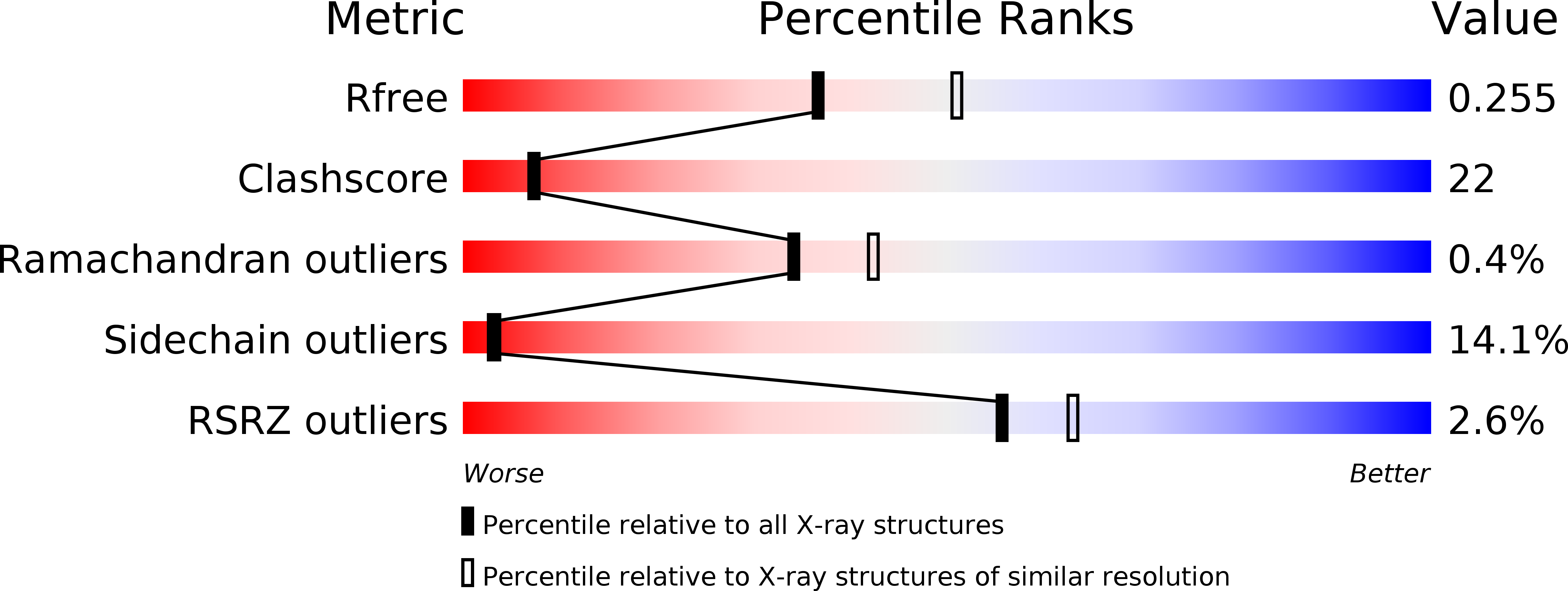
R-Value Free:
0.24
R-Value Work:
0.18
R-Value Observed:
0.18
Space Group:
P 21 21 21