1BE9 image
Deposition Date 1998-05-20
Release Date 1998-10-21
Last Version Date 2024-02-07
Entry Detail
PDB ID:
1BE9
Title:
THE THIRD PDZ DOMAIN FROM THE SYNAPTIC PROTEIN PSD-95 IN COMPLEX WITH A C-TERMINAL PEPTIDE DERIVED FROM CRIPT.
Biological Source:
Source Organism:
Host Organism:
Method Details:
Experimental Method:
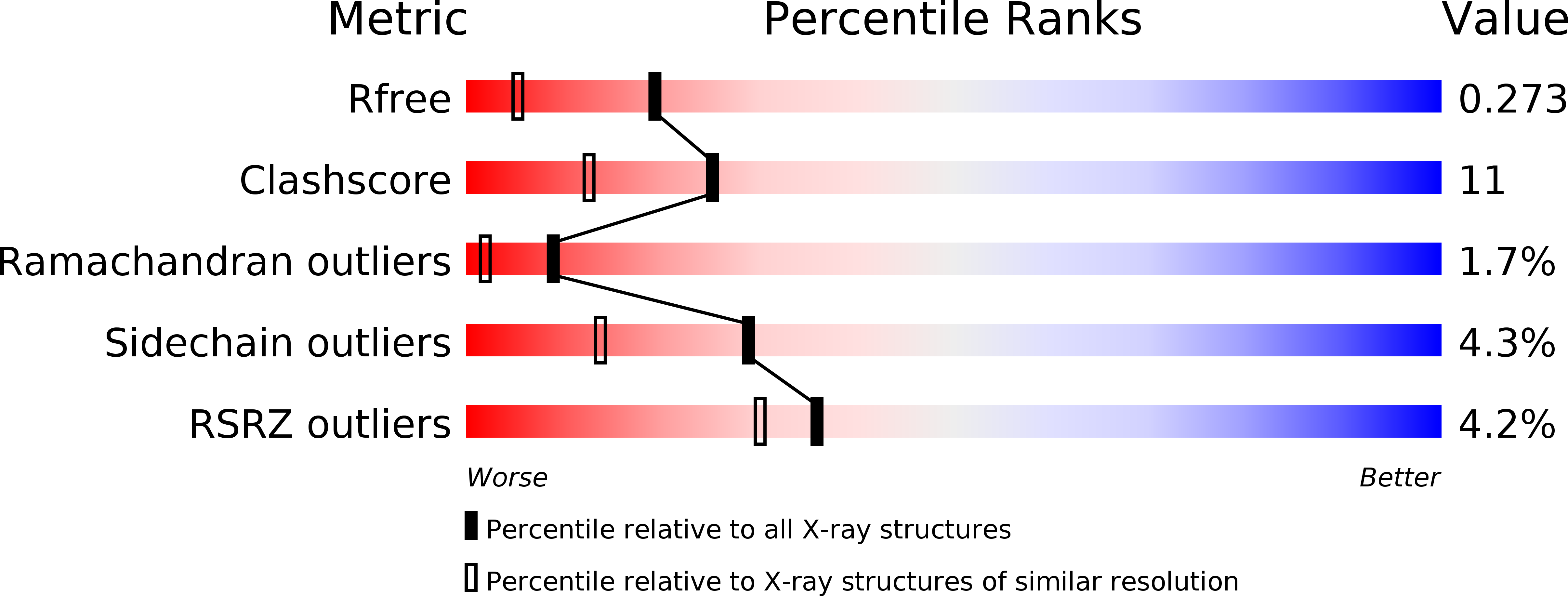
Resolution:
1.82 Å
R-Value Free:
0.27
R-Value Work:
0.20
Space Group:
P 41 3 2
Macromolecular Entities
Polymer Type:polypeptide(L)
Molecule:PSD-95
Gene (Uniprot):Dlg4
Chain IDs:A
Chain Length:119
Number of Molecules:1
Biological Source:Rattus norvegicus
Primary Citation
Crystal structures of a complexed and peptide-free membrane protein-binding domain: molecular basis of peptide recognition by PDZ.
Cell(Cambridge,Mass.) 85 1067 1076 (1996)
PMID: 8674113 DOI: 10.1016/S0092-8674(00)81307-0

Abstact

Modular PDZ domains, found in many cell junction-associated proteins, mediate the clustering of membrane ion channels by binding to their C-terminus. The X-ray crystallographic structures of the third PDZ domain from the synaptic protein PSD-95 in complex with and in the absence of its peptide ligand have been determined at 1.8 angstroms and 2.3 angstroms resolution, respectively. The structures reveal that a four-residue C-terminal stretch (X-Thr/Ser-X-Val-COO(-)) engages the PDZ domain through antiparallel main chain interactions with a beta sheet of the domain. Recognition of the terminal carboxylate group of the peptide is conferred by a cradle of main chain amides provided by a Gly-Leu-Gly-Phe loop as well as by an arginine side chain. Specific side chain interactions and a prominent hydrophobic pocket explain the selective recognition of the C-terminal consensus sequence.

Legend

Protein

Chemical

Disease

Primary Citation of related structures