1BDO image
Deposition Date 1995-11-21
Release Date 1996-08-01
Last Version Date 2024-10-23
Entry Detail
PDB ID:
1BDO
Keywords:
Title:
STRUCTURE OF THE BIOTINYL DOMAIN OF ACETYL-COENZYME A CARBOXYLASE DETERMINED BY MAD PHASING
Biological Source:
Source Organism:
Escherichia coli (Taxon ID: 562)
Host Organism:
Method Details:
Experimental Method:
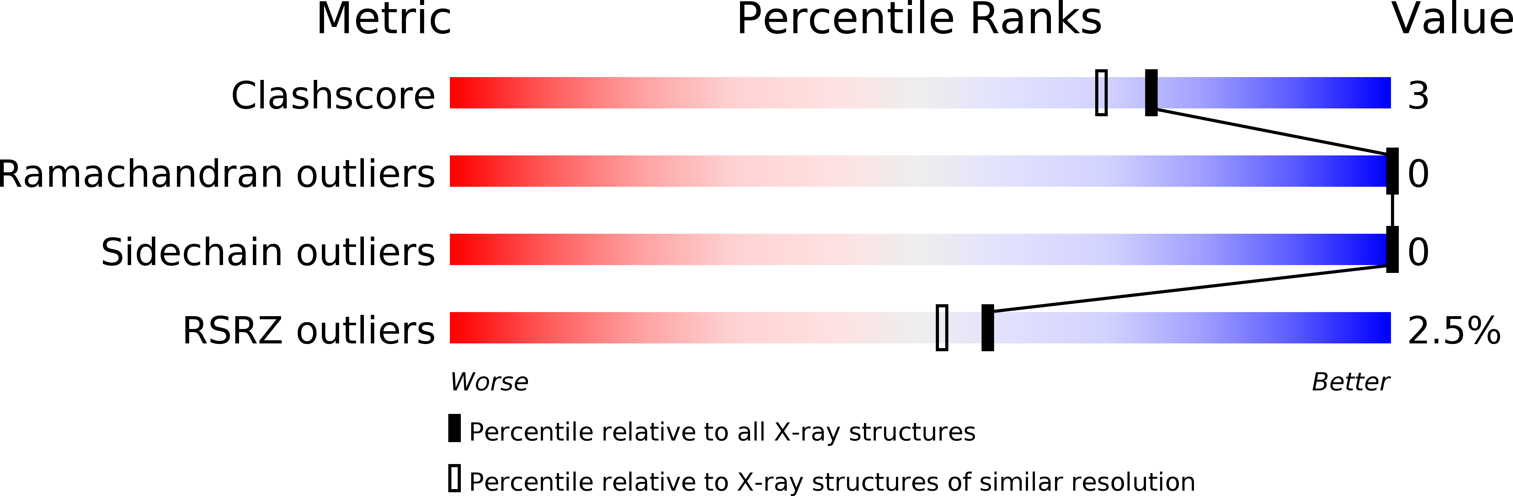
Resolution:
1.80 Å
R-Value Work:
0.18
R-Value Observed:
0.18
Space Group:
P 21 21 2
Macromolecular Entities
Polymer Type:polypeptide(L)
Molecule:ACETYL-COA CARBOXYLASE
Gene (Uniprot):accB
Chain IDs:A
Chain Length:80
Number of Molecules:1
Biological Source:Escherichia coli
Ligand Molecules
Primary Citation
Structure of the biotinyl domain of acetyl-coenzyme A carboxylase determined by MAD phasing.
Structure 3 1407 1419 (1995)
PMID: 8747466 DOI: 10.1016/S0969-2126(01)00277-5

Abstact

BACKGROUND Acetyl-coenzyme A carboxylase catalyzes the first committed step of fatty acid biosynthesis. Universally, this reaction involves three functional components all related to a carboxybiotinyl intermediate. A biotinyl domain shuttles its covalently attached biotin prosthetic group between the active sites of a biotin carboxylase and a carboxyl transferase. In Escherichia coli, the three components reside in separate subunits: a biotinyl domain is the functional portion of one of these, biotin carboxy carrier protein (BCCP). RESULTS We have expressed natural and selenomethionyl (Se-met) BCCP from E. coli as biotinylated recombinant proteins, proteolyzed them with subtilisin Carlsberg to produce the biotinyl domains BCCP and Se-met BCCPsc, determined the crystal structure of Se-met BCCPsc using a modified version of the multiwavelength anomalous diffraction (MAD) phasing protocol, and refined the structure for the natural BCCPsc at 1.8 A resolution. The structure may be described as a capped beta sandwich with quasi-dyad symmetry. Each half contains a characteristic hammerhead motif. The biotinylated lysin is located at a hairpin beta turn which connects the two symmetric halves of the molecule, and its biotinyl group interacts with a non-symmetric protrusion from the core. CONCLUSIONS This first crystal structure of a biotinyl domain helps to unravel the central role of such domains in reactions catalyzed by biotin-dependent carboxylases. The hammerhead structure observed twice in BCCPsc may be regarded as the basic structural motif of biotinyl and lipoyl domains of a superfamily of enzymes. The new MAD phasing techniques developed in the course of determining this structure enhance the power of the MAD method.

Legend

Protein

Chemical

Disease

Primary Citation of related structures