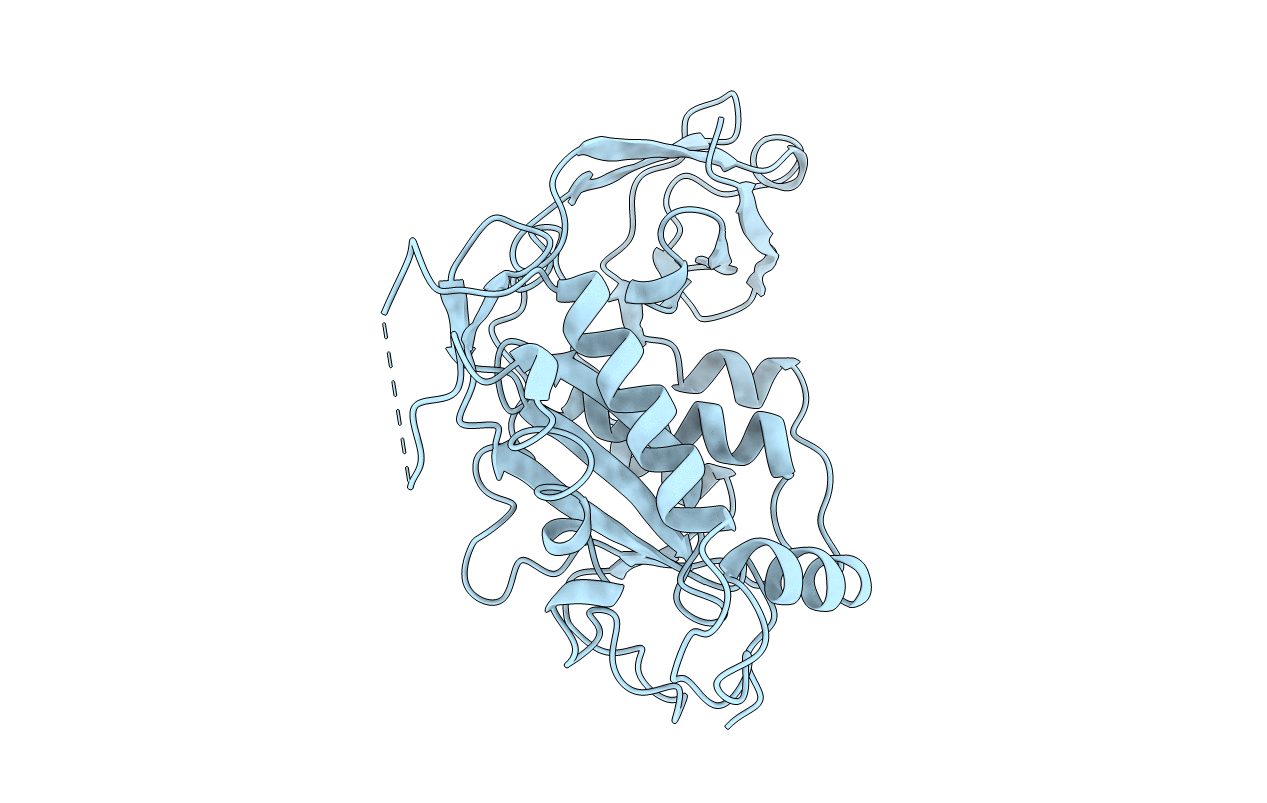
1BCO image
Deposition Date 1995-05-26
Release Date 1995-10-15
Last Version Date 2024-02-07
Entry Detail
PDB ID:
1BCO
Keywords:
Title:
BACTERIOPHAGE MU TRANSPOSASE CORE DOMAIN
Biological Source:
Source Organism:
Host Organism:
Method Details:
Experimental Method:
Resolution:
2.40 Å
R-Value Free:
0.26
R-Value Work:
0.19
R-Value Observed:
0.19
Space Group:
C 2 2 21
Macromolecular Entities
Structures with similar UniProt ID
Protein Blast
Polymer Type:polypeptide(L)
Molecule:BACTERIOPHAGE MU TRANSPOSASE
Gene (Uniprot):A
Chain IDs:A
Chain Length:327
Number of Molecules:1
Biological Source:Enterobacteria phage Mu
Primary Citation
Structure of the bacteriophage Mu transposase core: a common structural motif for DNA transposition and retroviral integration.
Cell(Cambridge,Mass.) 82 209 220 (1995)
PMID: 7628012 DOI: 10.1016/0092-8674(95)90308-9

Abstact

The crystal structure of the core domain of bacteriophage Mu transposase, MuA, has been determined at 2.4 A resolution. The first of two subdomains contains the active site and, despite very limited sequence homology, exhibits a striking similarity to the core domain of HIV-1 integrase, which carries out a similar set of biochemical reactions. It also exhibits more limited similarity to other nucleases, RNase H and RuvC. The second, a beta barrel, connects to the first subdomain through several contacts. Three independent determinations of the monomer structure from two crystal forms all show the active site held in a similar, apparently inactive configuration. The enzymatic activity of MuA is known to be activated by formation of a DNA-bound tetramer of the protein. We propose that the connections between the two subdomains may be involved in the cross-talk between the active site and the other domains of the transposase that controls the activity of the protein.

Legend

Protein

Chemical

Disease

Primary Citation of related structures