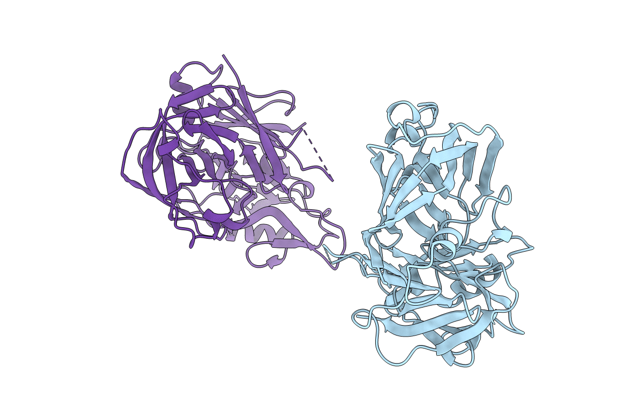
1BBS image
Entry Detail
PDB ID:
1BBS
Title:
X-RAY ANALYSES OF PEPTIDE INHIBITOR COMPLEXES DEFINE THE STRUCTURAL BASIS OF SPECIFICITY FOR HUMAN AND MOUSE RENINS
Biological Source:
Source Organism:
PDB Version:
Deposition Date:
1992-05-21
Release Date:
1994-01-31
Method Details:
Experimental Method:
Resolution:
2.80 Å
R-Value Observed:
0.19
Space Group:
P 21 3
Macromolecular Entities
Polymer Type:polypeptide(L)
Description:RENIN
Chain IDs:A, B
Chain Length:340
Number of Molecules:2
Biological Source:Homo sapiens
Ligand Molecules
Primary Citation
X-ray analyses of peptide-inhibitor complexes define the structural basis of specificity for human and mouse renins.
Nature 357 466 472 (1992)
PMID: 1608447 DOI: 10.1038/357466a0

Abstact

X-ray analyses have defined the three-dimensional structures of crystals of mouse and human renins complexed with peptide inhibitors at resolutions of 1.9 and 2.8 A, respectively. The exquisite specificity of renin arises partly from ordered loop regions at the periphery of the binding cleft. Although the pattern of main-chain hydrogen bonding in other aspartic proteinase inhibitor complexes is conserved in renins, differences in the positions of secondary structure elements (particularly helices) also lead to improved specificity in renins for angiotensinogen substrates.

Legend

Protein

Chemical

Disease

Primary Citation of related structures