1BBR image
Deposition Date 1992-04-27
Release Date 1994-01-31
Last Version Date 2024-10-30
Entry Detail
PDB ID:
1BBR
Keywords:
Title:
THE STRUCTURE OF RESIDUES 7-16 OF THE A ALPHA CHAIN OF HUMAN FIBRINOGEN BOUND TO BOVINE THROMBIN AT 2.3 ANGSTROMS RESOLUTION
Biological Source:
Source Organism:
Bos taurus (Taxon ID: 9913)
Homo sapiens (Taxon ID: 9606)
Method Details:
Experimental Method:
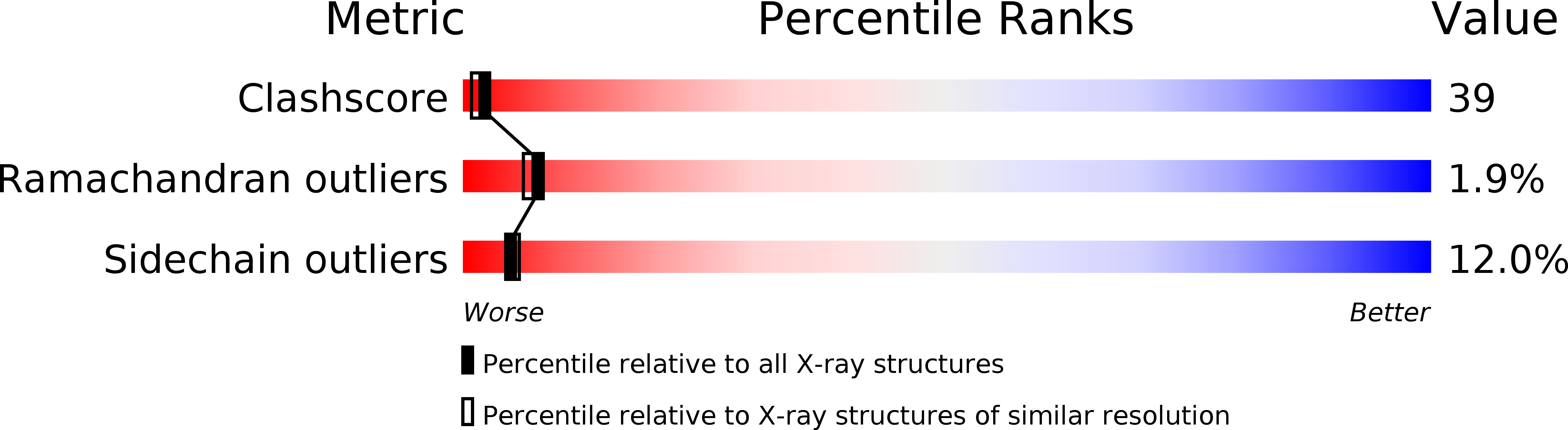
Resolution:
2.30 Å
R-Value Observed:
0.16
Space Group:
P 1 21 1
Macromolecular Entities
Structures with similar UniProt ID
Protein Blast
Polymer Type:polypeptide(L)
Molecule:EPSILON-THROMBIN
Gene (Uniprot):F2
Chain IDs:C (auth: E)
Chain Length:109
Number of Molecules:1
Biological Source:Bos taurus
Structures with similar UniProt ID
Protein Blast
Polymer Type:polypeptide(L)
Molecule:FIBRINOGEN ALPHA/ALPHA-E CHAIN PRECURSOR
Gene (Uniprot):FGA
Chain IDs:D (auth: F), G, J (auth: I)
Chain Length:11
Number of Molecules:3
Biological Source:Homo sapiens
Structures with similar UniProt ID
Protein Blast
Polymer Type:polypeptide(L)
Molecule:EPSILON-THROMBIN
Gene (Uniprot):F2
Chain IDs:B (auth: H)
Chain Length:150
Number of Molecules:1
Biological Source:Bos taurus
Structures with similar UniProt ID
Protein Blast
Polymer Type:polypeptide(L)
Molecule:EPSILON-THROMBIN
Gene (Uniprot):F2
Chain IDs:F (auth: K), I (auth: N)
Chain Length:259
Number of Molecules:2
Biological Source:Bos taurus
Structures with similar UniProt ID
Protein Blast
Polymer Type:polypeptide(L)
Molecule:EPSILON-THROMBIN
Gene (Uniprot):F2
Chain IDs:A (auth: L), E (auth: J), H (auth: M)
Chain Length:49
Number of Molecules:3
Biological Source:Bos taurus
Primary Citation
The structure of residues 7-16 of the A alpha-chain of human fibrinogen bound to bovine thrombin at 2.3-A resolution.
J.Biol.Chem. 267 7911 7920 (1992)
PMID: 1560020

Abstact

The tetradecapeptide Ac-D-F-L-A-E-G-G-G-V-R-G-P-R-V-OMe, which mimics residues 7f-20f of the A alpha-chain of human fibrinogen, has been co-crystallized with bovine thrombin from ammonium sulfate solutions in space group P2(1) with unit cell dimensions of a = 83.0 A, b = 89.4 A, c = 99.3 A, and beta = 106.6 degrees. Three crystallographically independent complexes were located in the asymmetric unit by molecular replacement using the native bovine thrombin structure as a model. The standard crystallographic R-factor is 0.167 at 2.3-A resolution. Excellent electron density could be traced for the decapeptide, beginning with Asp-7f and ending with Arg-16f in the active site of thrombin; the remaining 4 residues, which have been cleaved from the tetradecapeptide at the Arg-16f/Gly-17f bond, are not seen. Residues 7f-11f at the NH2 terminus of the peptide form a single turn of alpha-helix that is connected by Gly-12f, which has a positive phi angle, to an extended chain containing residues 13f-16f. The major specific interactions between the peptide and thrombin are 1) a hydrophobic cage formed by residues Tyr-60A, Trp-60D, Leu-99, Ile-174, Trp-215, Leu-9f, Gly-13f, and Val-15f that surrounds Phe-8f; 2) a hydrogen bond linking Phe-8f NH to Lys-97 O;3) a salt link between Glu-11f and Arg-173; 4) two antiparallel beta-sheet hydrogen bonds between Gly-14f and Gly-216; and 5) the insertion of Arg-16f into the specificity pocket. Binding of the peptide is accompanied by a considerable shift in two of the loops near the active site relative to human D-phenyl-L-prolyl-L-arginyl chloromethyl ketone (PPACK)-thrombin.

Legend

Protein

Chemical

Disease

Primary Citation of related structures
Feedback Form
Name
Email
Institute
Feedback