Deposition Date
1992-05-18
Release Date
1994-01-31
Last Version Date
2024-10-23
Entry Detail
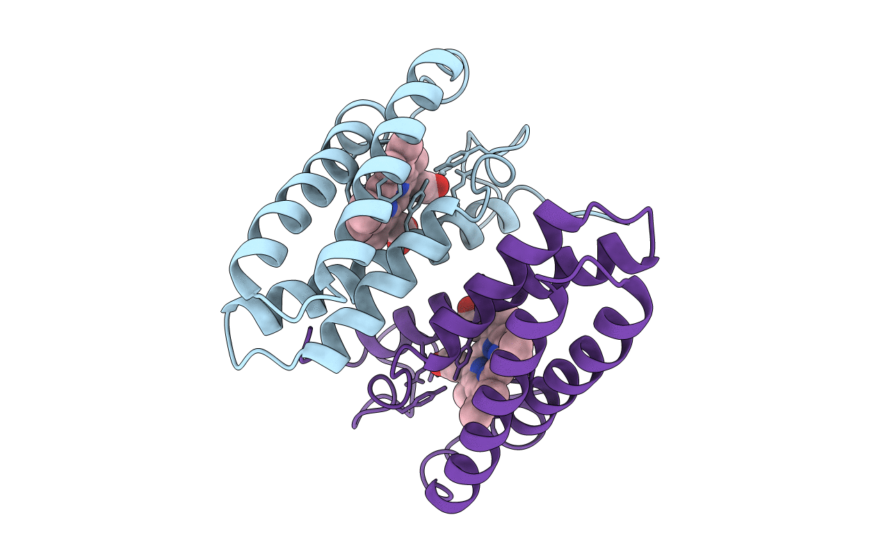
PDB ID:
1BBH
Keywords:
Title:
ATOMIC STRUCTURE OF A CYTOCHROME C' WITH AN UNUSUAL LIGAND-CONTROLLED DIMER DISSOCIATION AT 1.8 ANGSTROMS RESOLUTION
Biological Source:
Source Organism(s):
Allochromatium vinosum (Taxon ID: 1049)
Method Details:
Experimental Method:
Resolution:
1.80 Å
R-Value Work:
0.18
R-Value Observed:
0.18
Space Group:
P 21 21 21