1BA2 image
Deposition Date 1998-04-19
Release Date 1998-07-15
Last Version Date 2024-05-22
Entry Detail
PDB ID:
1BA2
Keywords:
Title:
D67R MUTANT OF D-RIBOSE-BINDING PROTEIN FROM ESCHERICHIA COLI
Biological Source:
Source Organism:
Host Organism:
Method Details:
Experimental Method:
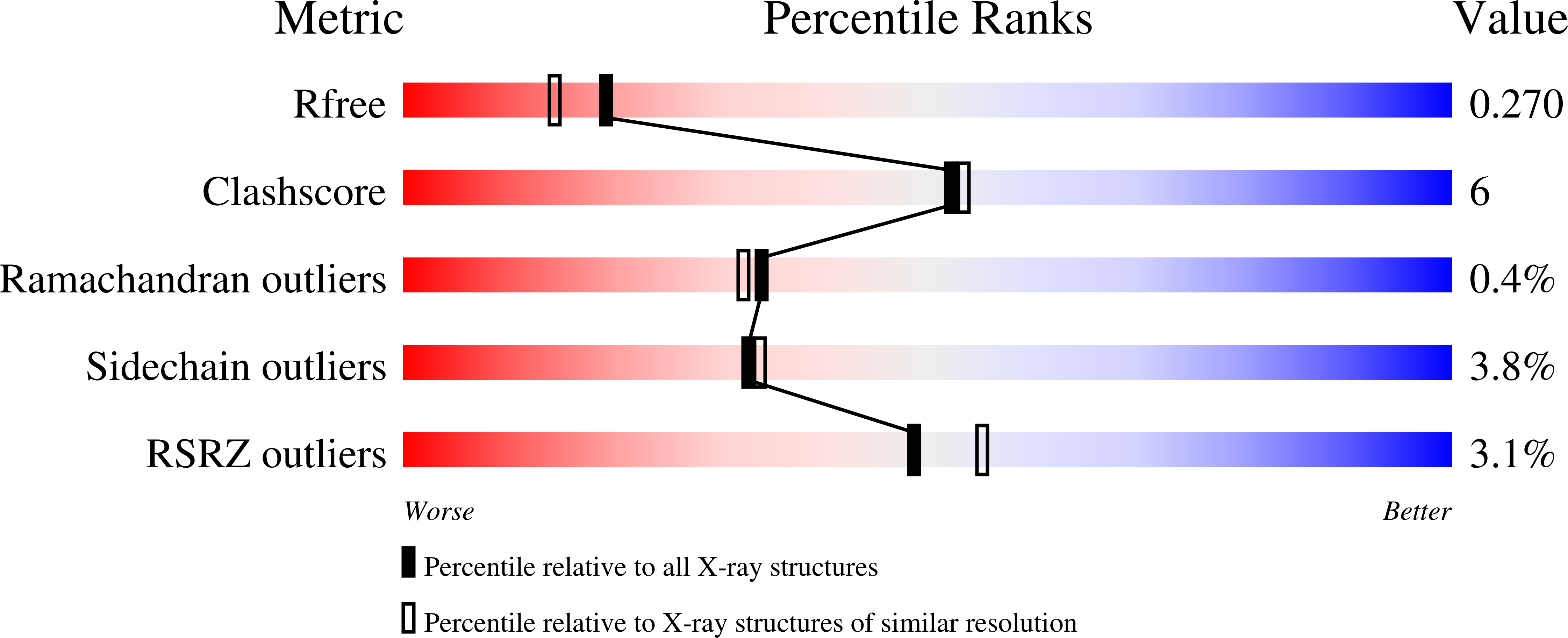
Resolution:
2.10 Å
R-Value Free:
0.27
R-Value Work:
0.19
Space Group:
P 21 21 21
Macromolecular Entities
Polymer Type:polypeptide(L)
Molecule:D-RIBOSE-BINDING PROTEIN
Gene (Uniprot):rbsB
Mutations:D67R
Chain IDs:A, B
Chain Length:271
Number of Molecules:2
Biological Source:Escherichia coli K12
Primary Citation
Multiple open forms of ribose-binding protein trace the path of its conformational change.
J.Mol.Biol. 279 651 664 (1998)
PMID: 9641984 DOI: 10.1006/jmbi.1998.1785

Abstact

Conformational changes are necessary for the function of bacterial periplasmic receptors in chemotaxis and transport. Such changes allow entry and exit of ligand, and enable the correct interaction of the ligand-bound proteins with the membrane components of each system. Three open, ligand-free forms of the Escherichia coli ribose-binding protein were observed here by X-ray crystallographic studies. They are opened by 43 degrees, 50 degrees and 64 degrees with respect to the ligand-bound protein reported previously. The three open forms are not distinct, but show a clear relationship to each other. All are the product of a similar opening motion, and are stabilized by a new, almost identical packing interface between the domains. The changes are generated by similar bond rotations, although some differences in the three hinge segments are needed to accommodate the various structural scenarios. Some local repacking also occurs as interdomain contacts are lost. The least open (43 degrees) form is probably the dominant one in solution under normal conditions, although a mixture of species seems likely. The open and closed forms have distinct surfaces in the regions known to be important in chemotaxis and transport, which will differentiate their interactions with the membrane components. It seems certain that the conformational path that links the forms described here is that followed during ligand retrieval, and in ligand release into the membrane-bound permease system.

Legend

Protein

Chemical

Disease

Primary Citation of related structures