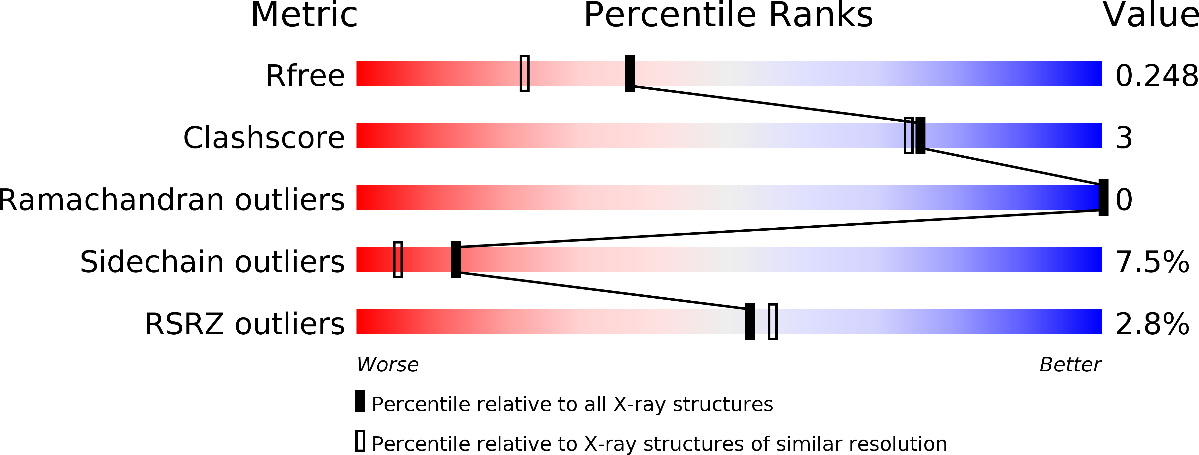
1B8R image
Entry Detail
PDB ID:
1B8R
Title:
PARVALBUMIN
Biological Source:
Source Organism:
Host Organism:
PDB Version:
Deposition Date:
1999-02-02
Release Date:
1999-02-09
Method Details:
Experimental Method:
Resolution:
1.90 Å
R-Value Free:
0.27
R-Value Work:
0.17
Space Group:
C 1 2 1
Macromolecular Entities
Polymer Type:polypeptide(L)
Description:PROTEIN (PARVALBUMIN)
Mutations:F102W
Chain IDs:A
Chain Length:108
Number of Molecules:1
Biological Source:Cyprinus carpio
Ligand Molecules
Primary Citation
Metal-ion affinity and specificity in EF-hand proteins: coordination geometry and domain plasticity in parvalbumin.
Structure Fold.Des. 7 1269 1278 (1999)
PMID: 10545326 DOI: 10.1016/S0969-2126(00)80060-X

Abstact

BACKGROUND: The EF-hand family is a large set of Ca(2+)-binding proteins that contain characteristic helix-loop-helix binding motifs that are highly conserved in sequence. Members of this family include parvalbumin and many prominent regulatory proteins such as calmodulin and troponin C. EF-hand proteins are involved in a variety of physiological processes including cell-cycle regulation, second messenger production, muscle contraction, microtubule organization and vision. RESULTS: We have determined the structures of parvalbumin mutants designed to explore the role of the last coordinating residue of the Ca(2+)-binding loop. An E101D substitution has been made in the parvalbumin EF site. The substitution decreases the Ca(2+)-binding affinity 100-fold and increases the Mg(2+)-binding affinity 10-fold. Both the Ca(2+)- and Mg(2+)-bound structures have been determined, and a structural basis has been proposed for the metal-ion-binding properties. CONCLUSIONS: The E101D mutation does not affect the Mg(2+) coordination geometry of the binding loop, but it does pull the F helix 1.1 A towards the loop. The E101D-Ca(2+) structure reveals that this mutant cannot obtain the sevenfold coordination preferred by Ca(2+), presumably because of strain limits imposed by tertiary structure. Analysis of these results relative to previously reported structural information supports a model wherein the characteristics of the last coordinating residue and the plasticity of the Ca(2+)-binding loop delimit the allowable geometries for the coordinating sphere.

Legend

Protein

Chemical

Disease

Primary Citation of related structures