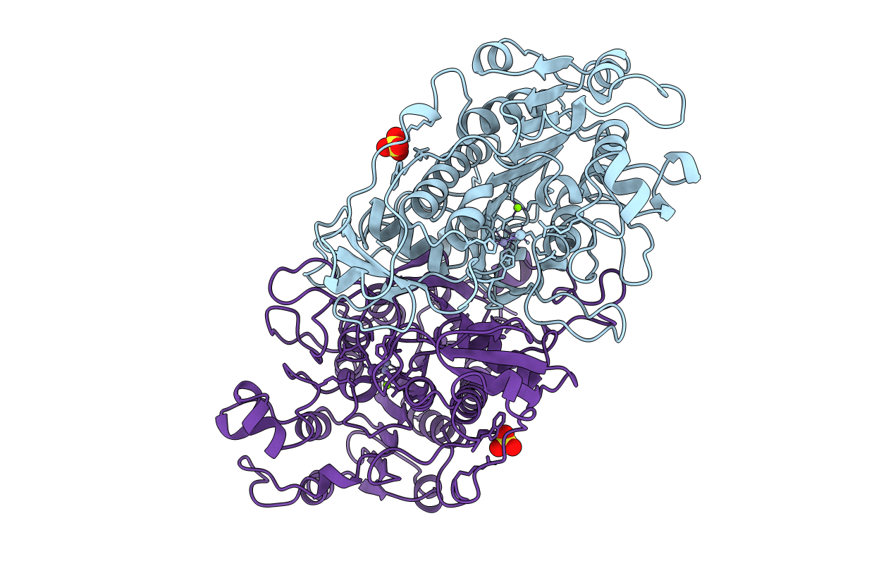
1B8J image
Deposition Date 1999-02-01
Release Date 1999-02-18
Last Version Date 2024-10-16
Entry Detail
PDB ID:
1B8J
Title:
ALKALINE PHOSPHATASE COMPLEXED WITH VANADATE
Biological Source:
Source Organism:
Escherichia coli (Taxon ID: 562)
Host Organism:
Method Details:
Experimental Method:
Resolution:
1.90 Å
R-Value Free:
0.19
R-Value Work:
0.17
Space Group:
I 2 2 2
Macromolecular Entities
Structures with similar UniProt ID
Protein Blast
Polymer Type:polypeptide(L)
Molecule:PROTEIN (ALKALINE PHOSPHATASE)
Gene (Uniprot):phoA
Chain IDs:A, B
Chain Length:449
Number of Molecules:2
Biological Source:Escherichia coli
Modified Residue
Compound ID Chain ID Parent Comp ID Details 2D Image
SVA A SER SERINE VANADATE
Primary Citation
A model of the transition state in the alkaline phosphatase reaction.
J.Biol.Chem. 274 8351 8354 (1999)
PMID: 10085061 DOI: 10.1074/jbc.274.13.8351

Abstact

A high resolution crystal structure of Escherichia coli alkaline phosphatase in the presence of vanadate has been refined to 1.9 A resolution. The vanadate ion takes on a trigonal bipyramidal geometry and is covalently bound by the active site serine nucleophile. A coordinated water molecule occupies the axial position opposite the serine nucleophile, whereas the equatorial oxygen atoms of the vanadate ion are stabilized by interactions with both Arg-166 and the zinc metal ions of the active site. This structural complex supports the in-line displacement mechanism of phosphomonoester hydrolysis by alkaline phosphatase and provides a model for the proposed transition state in the enzyme-catalyzed reaction.

Legend

Protein

Chemical

Disease

Primary Citation of related structures