1B6C image
Deposition Date 1999-01-13
Release Date 1999-06-15
Last Version Date 2024-02-07
Entry Detail
PDB ID:
1B6C
Title:
CRYSTAL STRUCTURE OF THE CYTOPLASMIC DOMAIN OF THE TYPE I TGF-BETA RECEPTOR IN COMPLEX WITH FKBP12
Biological Source:
Source Organism:
Homo sapiens (Taxon ID: 9606)
Host Organism:
Method Details:
Experimental Method:
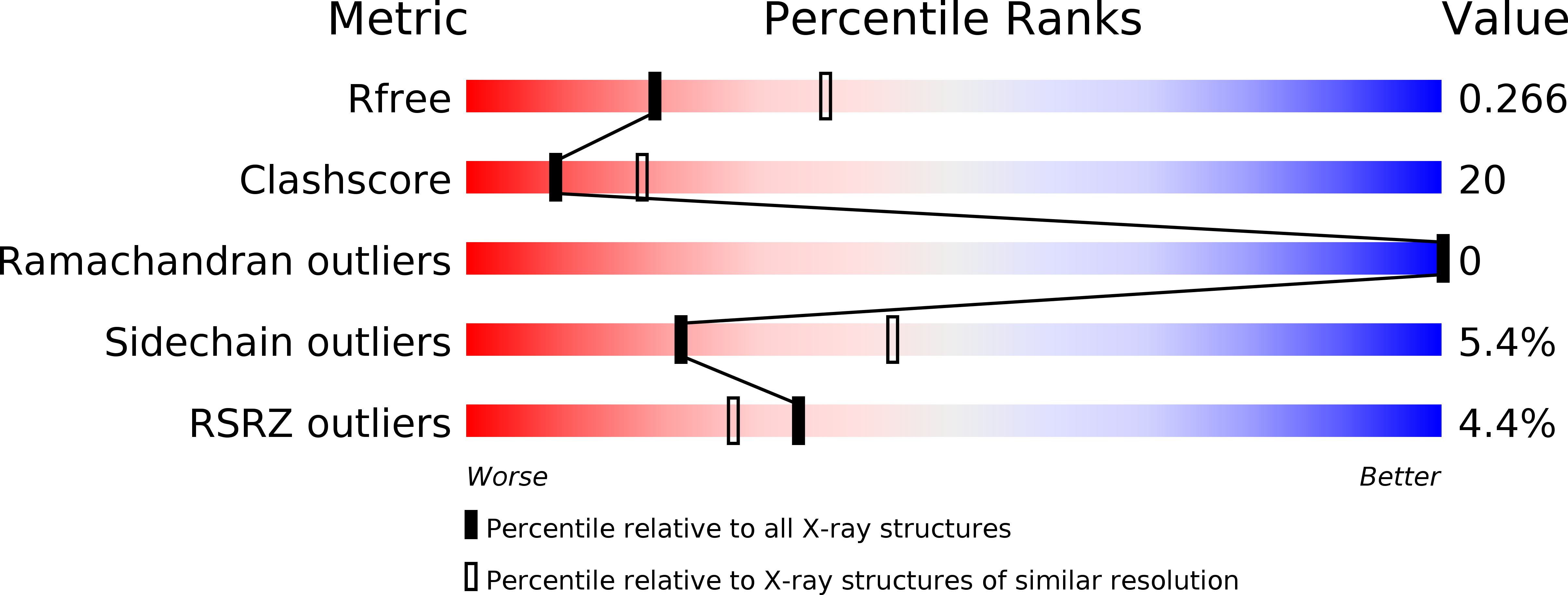
Resolution:
2.60 Å
R-Value Free:
0.26
R-Value Work:
0.24
R-Value Observed:
0.24
Space Group:
P 1
Macromolecular Entities
Polymer Type:polypeptide(L)
Molecule:FK506-BINDING PROTEIN
Gene (Uniprot):FKBP1A
Chain IDs:A, C, E, G
Chain Length:107
Number of Molecules:4
Biological Source:Homo sapiens
Polymer Type:polypeptide(L)
Molecule:TGF-B SUPERFAMILY RECEPTOR TYPE I
Gene (Uniprot):TGFBR1
Chain IDs:B, D, F, H
Chain Length:342
Number of Molecules:4
Biological Source:Homo sapiens
Ligand Molecules
Primary Citation
Crystal structure of the cytoplasmic domain of the type I TGF beta receptor in complex with FKBP12.
Cell(Cambridge,Mass.) 96 425 436 (1999)
PMID: 10025408 DOI: 10.1016/S0092-8674(00)80555-3

Abstact

Activation of the type I TGFbeta receptor (TbetaR-I) requires phosphorylation of a regulatory segment known as the GS region, located upstream of the serine/threonine kinase domain in the cytoplasmic portion of the receptor. The crystal structure of a fragment of unphosphorylated TbetaR-I, containing both the GS region and the catalytic domain, has been determined in complex with the FK506-binding protein FKBP12. TbetaR-I adopts an inactive conformation that is maintained by the unphosphorylated GS region. FKBP12 binds to the GS region of the receptor, capping the TbetaR-II phosphorylation sites and further stabilizing the inactive conformation of TbetaR-I. Certain structural features at the catalytic center of TbetaR-I are characteristic of tyrosine kinases rather than Ser/Thr kinases.

Legend

Protein

Chemical

Disease

Primary Citation of related structures