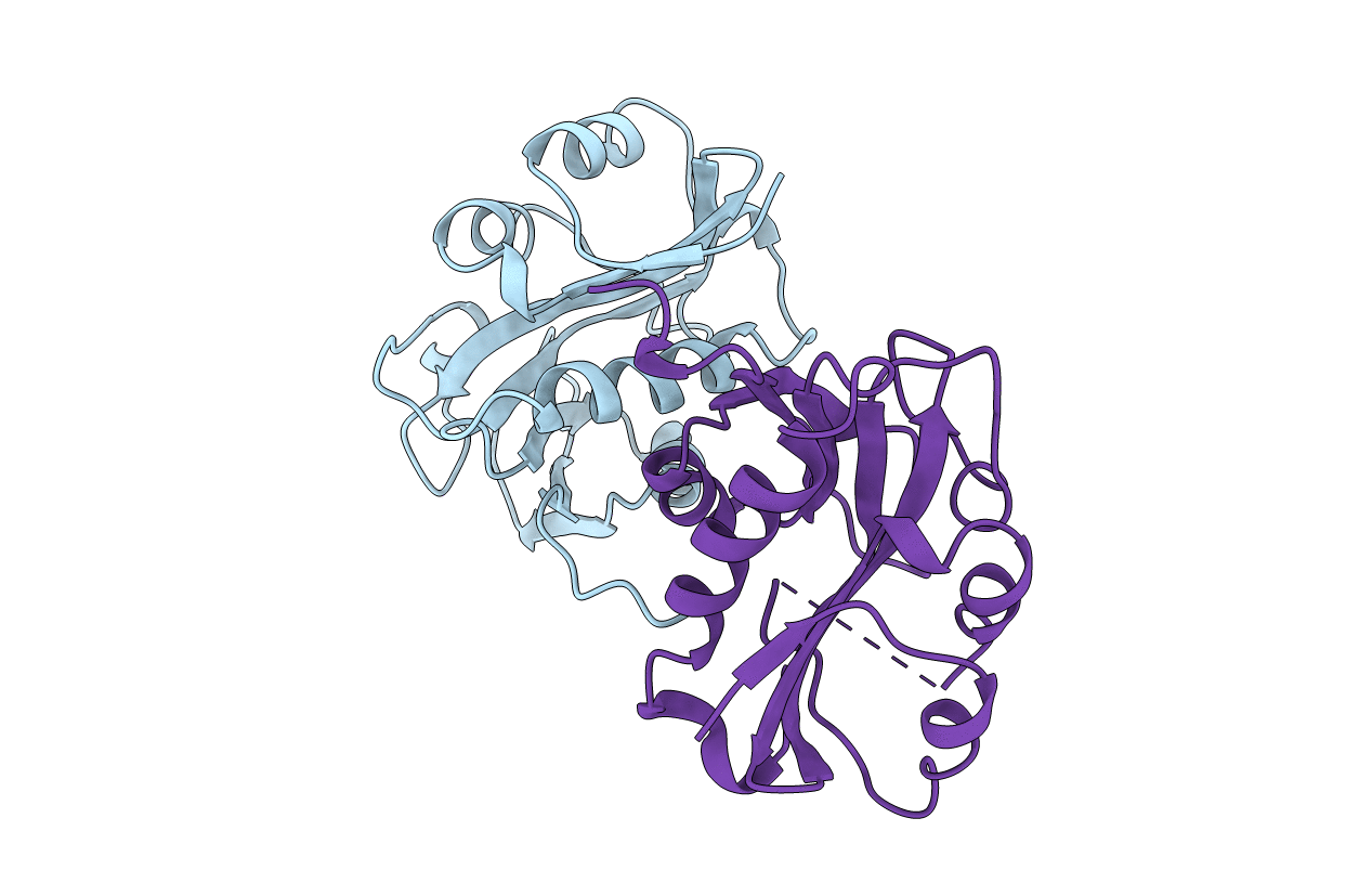
1B6B image
Deposition Date 1999-01-13
Release Date 2000-01-14
Last Version Date 2023-12-27
Entry Detail
PDB ID:
1B6B
Keywords:
Title:
MELATONIN BIOSYNTHESIS: THE STRUCTURE OF SEROTONIN N-ACETYLTRANSFERASE AT 2.5 A RESOLUTION SUGGESTS A CATALYTIC MECHANISM
Biological Source:
Source Organism:
Ovis aries (Taxon ID: 9940)
Host Organism:
Method Details:
Experimental Method:
Resolution:
2.50 Å
R-Value Free:
0.28
R-Value Work:
0.21
R-Value Observed:
0.21
Space Group:
P 61
Macromolecular Entities
Structures with similar UniProt ID
Protein Blast
Polymer Type:polypeptide(L)
Molecule:PROTEIN (ARYLALKYLAMINE N-ACETYLTRANSFERASE)
Gene (Uniprot):AANAT
Chain IDs:A, B
Chain Length:174
Number of Molecules:2
Biological Source:Ovis aries
Primary Citation
Melatonin biosynthesis: the structure of serotonin N-acetyltransferase at 2.5 A resolution suggests a catalytic mechanism.
Mol.Cell 3 23 32 (1999)
PMID: 10024876 DOI: 10.1016/S1097-2765(00)80171-9

Abstact

Conversion of serotonin to N-acetylserotonin, the precursor of the circadian neurohormone melatonin, is catalyzed by serotonin N-acetyltransferase (AANAT) in a reaction requiring acetyl coenzyme A (AcCoA). AANAT is a globular protein consisting of an eight-stranded beta sheet flanked by five alpha helices; a conserved motif in the center of the beta sheet forms the cofactor binding site. Three polypeptide loops converge above the AcCoA binding site, creating a hydrophobic funnel leading toward the cofactor and serotonin binding sites in the protein interior. Two conserved histidines not found in other NATs are located at the bottom of the funnel in the active site, suggesting a catalytic mechanism for acetylation involving imidazole groups acting as general acid/base catalysts.

Legend

Protein

Chemical

Disease

Primary Citation of related structures
Feedback Form
Name
Email
Institute
Feedback