Deposition Date
1999-01-12
Release Date
2000-01-07
Last Version Date
2023-08-09
Entry Detail
PDB ID:
1B57
Keywords:
Title:
CLASS II FRUCTOSE-1,6-BISPHOSPHATE ALDOLASE IN COMPLEX WITH PHOSPHOGLYCOLOHYDROXAMATE
Biological Source:
Source Organism(s):
Escherichia coli (Taxon ID: 562)
Expression System(s):
Method Details:
Experimental Method:
Resolution:
2.00 Å
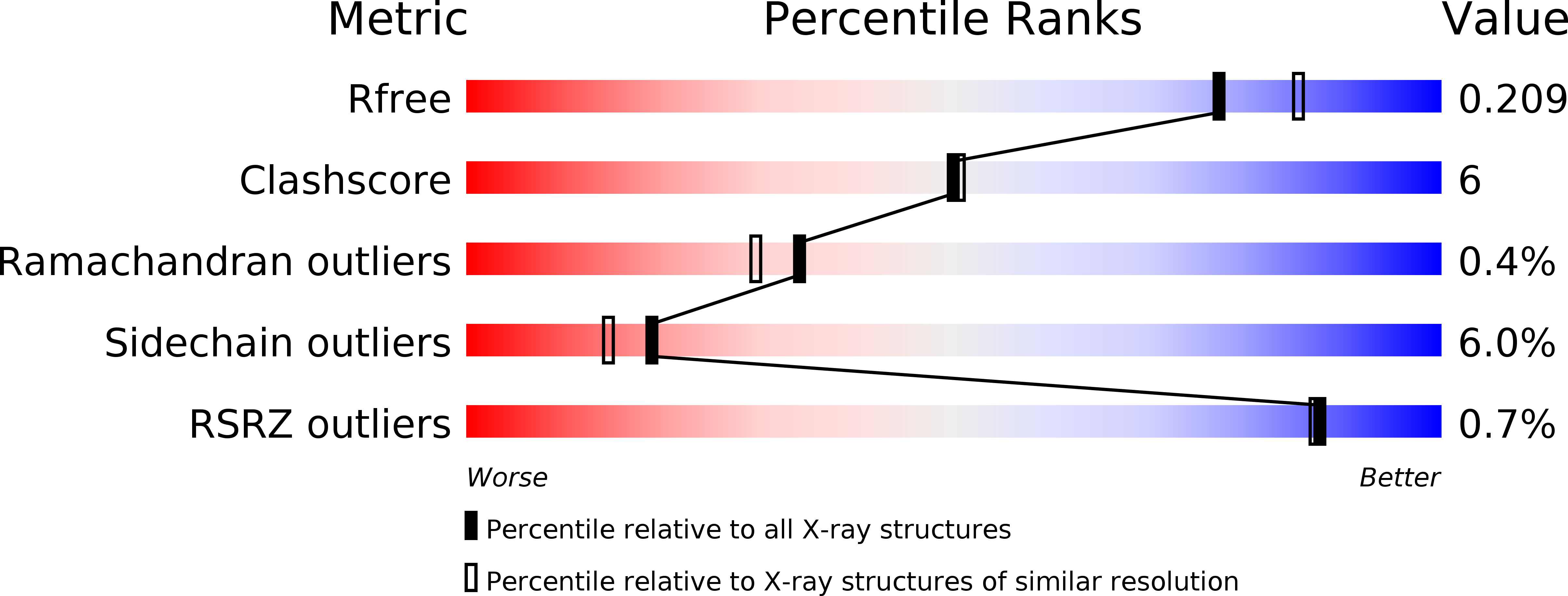
R-Value Free:
0.23
R-Value Work:
0.19
Space Group:
P 31 2 1