1B4G image
Entry Detail
PDB ID:
1B4G
Title:
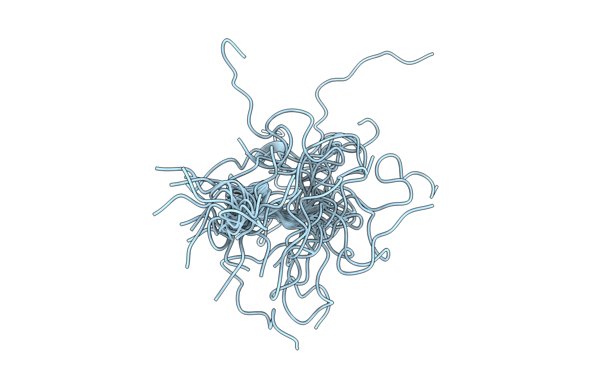
CONTROL OF K+ CHANNEL GATING BY PROTEIN PHOSPHORYLATION: STRUCTURAL SWITCHES OF THE INACTIVATION GATE, NMR, 22 STRUCTURES
Biological Source:
Source Organism:
PDB Version:
Deposition Date:
1998-12-22
Release Date:
1999-04-27
Method Details:
Experimental Method:
Conformers Submitted:
22
Macromolecular Entities
Polymer Type:polypeptide(L)
Description:POTASSIUM CHANNEL
Chain IDs:A
Chain Length:30
Number of Molecules:1
Biological Source:Homo sapiens
Modified Residue
Compound ID Chain ID Parent Comp ID Details 2D Image
SEP A SER PHOSPHOSERINE
Ligand Molecules
Primary Citation
Control of K+ channel gating by protein phosphorylation: structural switches of the inactivation gate.
Nat.Struct.Biol. 6 146 150 (1999)
PMID: 10048926 DOI: 10.1038/5833

Abstact

Fast N-type inactivation of voltage-dependent potassium (Kv) channels controls membrane excitability and signal propagation in central neurons and occurs by a 'ball-and-chain'-type mechanism. In this mechanism an N-terminal protein domain (inactivation gate) occludes the pore from the cytoplasmic side. In Kv3.4 channels, inactivation is not fixed but is dynamically regulated by protein phosphorylation. Phosphorylation of several identified serine residues on the inactivation gate leads to reduction or removal of fast inactivation. Here, we investigate the structure-function basis of this phospho-regulation with nuclear magnetic resonance (NMR) spectroscopy and patch-clamp recordings using synthetic inactivation domains (ID). The dephosphorylated ID exhibited compact structure and displayed high-affinity binding to its receptor. Phosphorylation of serine residues in the N- or C-terminal half of the ID resulted in a loss of overall structural stability. However, depending on the residue(s) phosphorylated, distinct structural elements remained stable. These structural changes correlate with the distinct changes in binding and unbinding kinetics underlying the reduced inactivation potency of phosphorylated IDs.

Legend

Protein

Chemical

Disease

Primary Citation of related structures