
Deposition Date
1998-12-14
Release Date
1999-12-15
Last Version Date
2023-12-27
Entry Detail
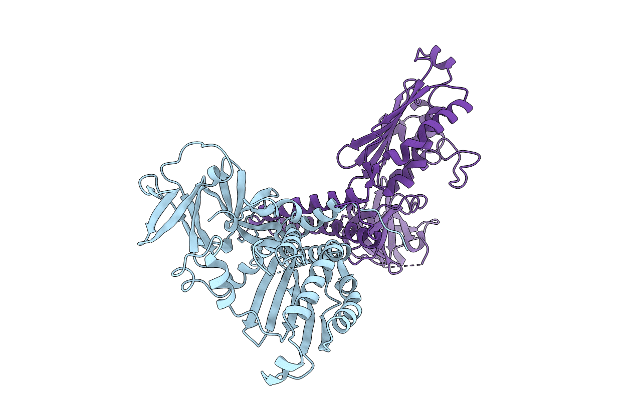
PDB ID:
1B3Q
Keywords:
Title:
CRYSTAL STRUCTURE OF CHEA-289, A SIGNAL TRANSDUCING HISTIDINE KINASE
Biological Source:
Source Organism(s):
Thermotoga maritima (Taxon ID: 2336)
Expression System(s):
Method Details:
Experimental Method:
Resolution:
2.60 Å
R-Value Free:
0.28
R-Value Work:
0.21
Space Group:
P 1 21 1
