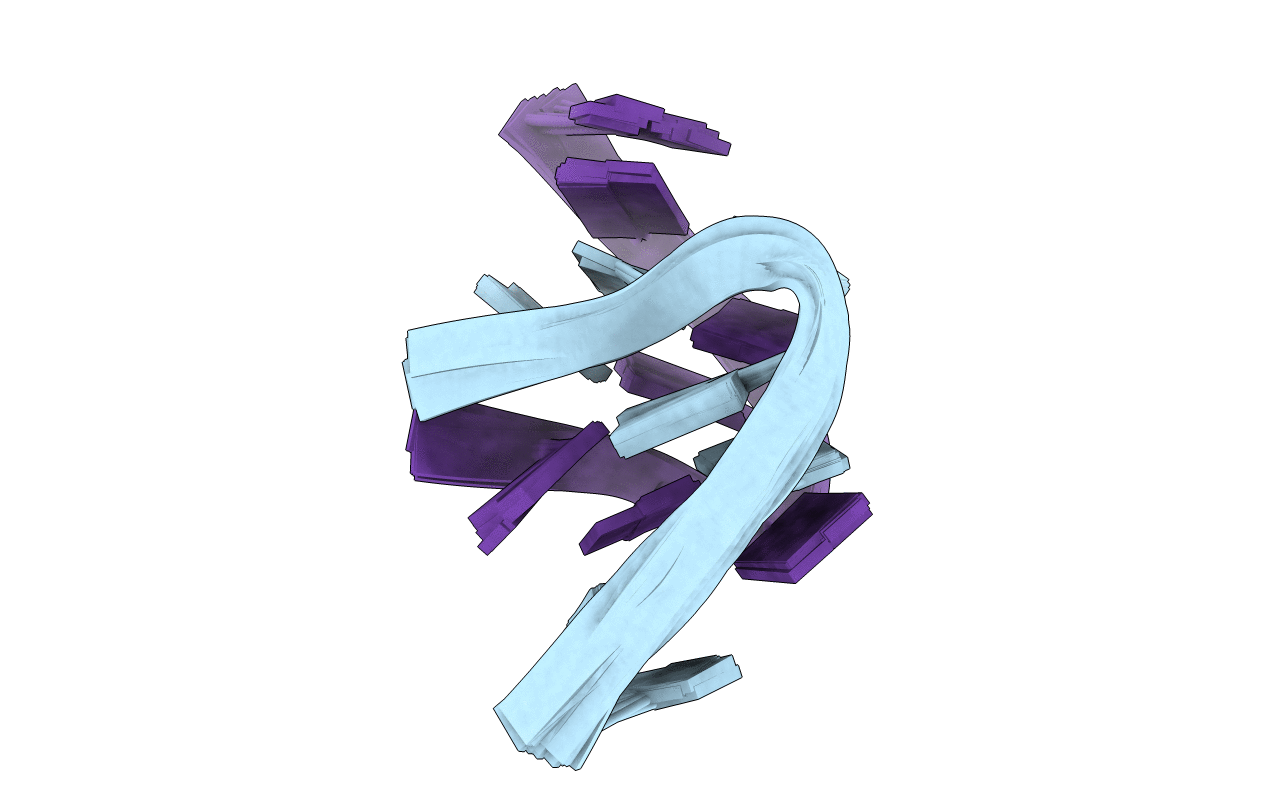
1B3P image
Deposition Date 1998-12-14
Release Date 1999-08-31
Last Version Date 2023-12-27
Entry Detail
PDB ID:
1B3P
Keywords:
Title:
5'-D(*GP*GP*AP*GP*GP*AP*T)-3'
Biological Source:
Source Organism:
(Taxon ID: ) (Taxon ID: )
Method Details:
Experimental Method:
Conformers Calculated:
100
Conformers Submitted:
15
Selection Criteria:
ACCEPTABLE COVALENT GEOMETRY, LOW DISTANCE RESTRAINTS VIIOLATIONS AND FAVORABLE NON-BONDED ENERGY VALUES
Macromolecular Entities
Polymer Type:polydeoxyribonucleotide
Molecule:DNA (5'-D(*GP*GP*AP*GP*GP*AP*T)-3')
Chain IDs:A, B
Chain Length:7
Number of Molecules:2
Biological Source:
Ligand Molecules
Primary Citation
Interlocked mismatch-aligned arrowhead DNA motifs.
Structure Fold.Des. 7 803 815 (1999)
PMID: 10425682 DOI: 10.1016/S0969-2126(99)80104-X

Abstact

BACKGROUND: Triplet repeat sequences are of considerable biological importance as the expansion of such tandem arrays can lead to the onset of a range of human diseases. Such sequences can self-pair via mismatch alignments to form higher order structures that have the potential to cause replication blocks, followed by strand slippage and sequence expansion. The all-purine d(GGA)n triplet repeat sequence is of particular interest because purines can align via G.G, A.A and G.A mismatch formation. RESULTS: We have solved the structure of the uniformly 13C,15N-labeled d(G1-G2-A3-G4-G5-A6-T7) sequence in 10 mM Na+ solution. This sequence adopts a novel twofold-symmetric duplex fold where interlocked V-shaped arrowhead motifs are aligned solely via interstrand G1.G4, G2.G5 and A3.A6 mismatch formation. The tip of the arrowhead motif is centered about the p-A3-p step, and symmetry-related local parallel-stranded duplex domains are formed by the G1-G2-A3 and G4-G5-A6 segments of partner strands. CONCLUSIONS: The purine-rich (GGA)n triplet repeat sequence is dispersed throughout the eukaryotic genome. Several features of the arrowhead duplex motif for the (GGA)2 triplet repeat provide a unique scaffold for molecular recognition. These include the large localized bend in the sugar-phosphate backbones, the segmental parallel-stranded alignment of strands and the exposure of the Watson-Crick edges of several mismatched bases.

Legend

Protein

Chemical

Disease

Primary Citation of related structures