1B3N image
Deposition Date 1998-12-14
Release Date 1999-04-06
Last Version Date 2024-10-16
Entry Detail
PDB ID:
1B3N
Title:
BETA-KETOACYL CARRIER PROTEIN SYNTHASE AS A DRUG TARGET, IMPLICATIONS FROM THE CRYSTAL STRUCTURE OF A COMPLEX WITH THE INHIBITOR CERULENIN.
Biological Source:
Source Organism:
Host Organism:
Method Details:
Experimental Method:
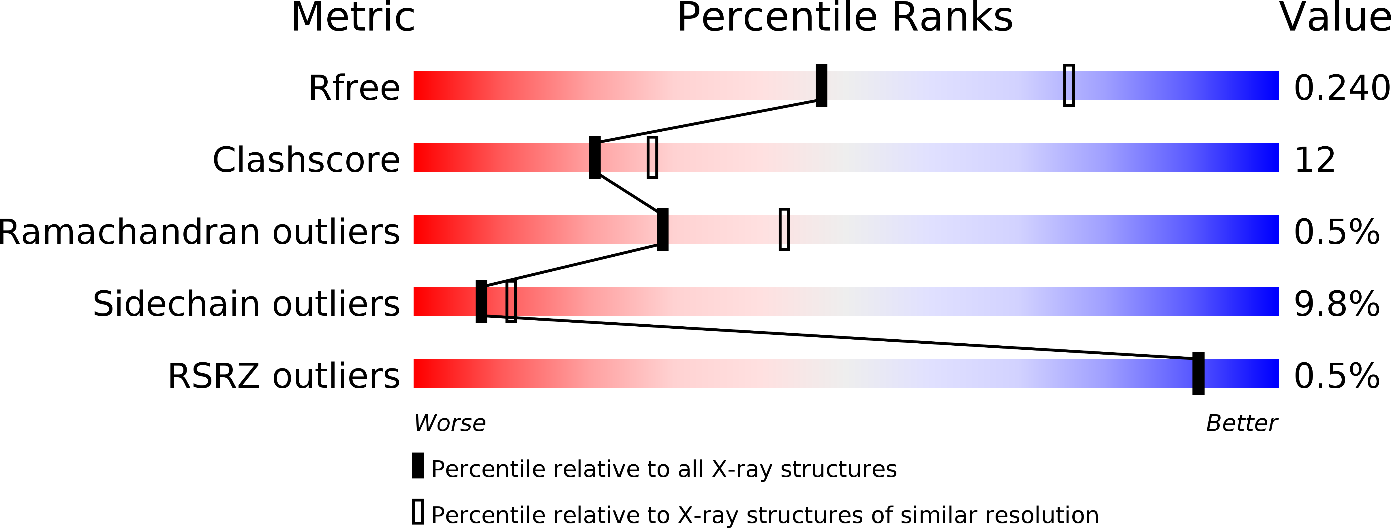
Resolution:
2.65 Å
R-Value Free:
0.27
R-Value Work:
0.21
Space Group:
P 31 2 1
Macromolecular Entities
Structures with similar UniProt ID
Protein Blast
Polymer Type:polypeptide(L)
Molecule:PROTEIN (KETOACYL ACYL CARRIER PROTEIN SYNTHASE 2)
Gene (Uniprot):fabF
Chain IDs:A
Chain Length:412
Number of Molecules:1
Biological Source:Escherichia coli
Ligand Molecules
Primary Citation
Structure of the complex between the antibiotic cerulenin and its target, beta-ketoacyl-acyl carrier protein synthase.
J.Biol.Chem. 274 6031 6034 (1999)
PMID: 10037680 DOI: 10.1074/jbc.274.10.6031

Abstact

In the biosynthesis of fatty acids, the beta-ketoacyl-acyl carrier protein (ACP) synthases catalyze chain elongation by the addition of two-carbon units derived from malonyl-ACP to an acyl group bound to either ACP or CoA. The enzyme is a possible drug target for treatment of certain cancers and for tuberculosis. The crystal structure of the complex of the enzyme from Escherichia coli, and the fungal mycotoxin cerulenin reveals that the inhibitor is bound in a hydrophobic pocket formed at the dimer interface. Cerulenin is covalently attached to the active site cysteine through its C2 carbon atom. The fit of the inhibitor to the active site is not optimal, and there is thus room for improvement through structure based design.

Legend

Protein

Chemical

Disease

Primary Citation of related structures
Feedback Form
Name
Email
Institute
Feedback