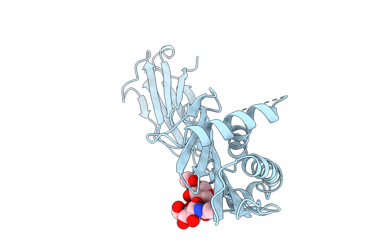
1B3J image
Deposition Date 1998-12-11
Release Date 1999-07-09
Last Version Date 2024-10-09
Entry Detail
PDB ID:
1B3J
Keywords:
Title:
STRUCTURE OF THE MHC CLASS I HOMOLOG MIC-A, A GAMMADELTA T CELL LIGAND
Biological Source:
Source Organism:
Homo sapiens (Taxon ID: 9606)
Host Organism:
Method Details:
Experimental Method:
Resolution:
3.00 Å
R-Value Free:
0.28
R-Value Work:
0.24
R-Value Observed:
0.24
Space Group:
F 41 3 2
Macromolecular Entities
Structures with similar UniProt ID
Protein Blast
Polymer Type:polypeptide(L)
Molecule:MHC CLASS I HOMOLOG MIC-A
Chain IDs:A
Chain Length:274
Number of Molecules:1
Biological Source:Homo sapiens
Modified Residue
Compound ID Chain ID Parent Comp ID Details 2D Image
ASN A ASN GLYCOSYLATION SITE
Primary Citation
Crystal structure of the MHC class I homolog MIC-A, a gammadelta T cell ligand.
Immunity 10 577 584 (1999)
PMID: 10367903 DOI: 10.1016/S1074-7613(00)80057-6

Abstact

The major histocompatibility complex (MHC) class I homolog MIC-A functions as a stress-inducible antigen that is recognized by a subset of gammadelta T cells independent of beta2-microglobulin and bound peptides. Its crystal structure reveals a dramatically altered MHC class I fold, both in detail and overall domain organization. The only remnant of a peptide-binding groove is a small cavity formed as the result of disordering a large section of one of the groove-defining helices. Loss of beta2-microglobulin binding is due to a restructuring of the interaction interfaces. Structural mapping of sequence variation suggests potential receptor binding sites on the underside of the platform on the side opposite of the surface recognized by alphabeta T cell receptors on MHC class I-peptide complexes.

Legend

Protein

Chemical

Disease

Primary Citation of related structures