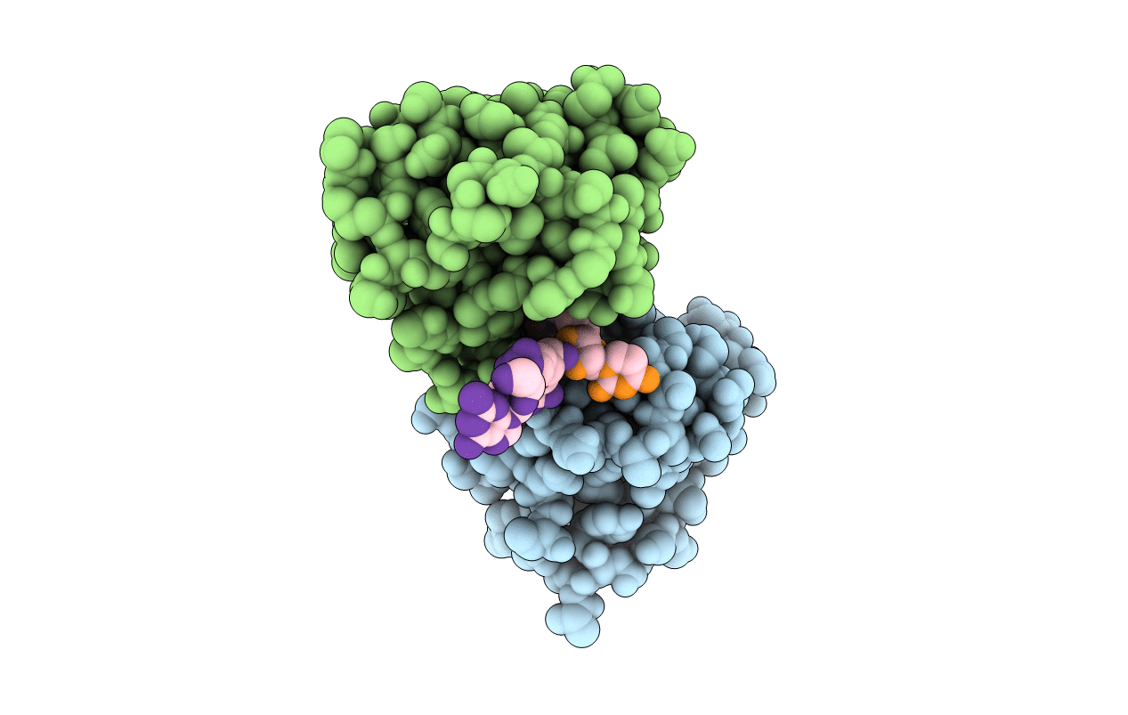
1B2M image
Deposition Date 1998-11-27
Release Date 1999-03-25
Last Version Date 2024-10-30
Entry Detail
PDB ID:
1B2M
Keywords:
Title:
THREE-DIMENSIONAL STRUCTURE OF RIBONULCEASE T1 COMPLEXED WITH AN ISOSTERIC PHOSPHONATE ANALOGUE OF GPU: ALTERNATE SUBSTRATE BINDING MODES AND CATALYSIS.
Biological Source:
Source Organism:
Method Details:
Experimental Method:
Resolution:
2.00 Å
R-Value Free:
0.25
R-Value Work:
0.18
R-Value Observed:
0.18
Space Group:
P 21 21 21
Macromolecular Entities
Structures with similar UniProt ID
Protein Blast
Polymer Type:polypeptide(L)
Molecule:RIBONUCLEASE T1
Gene (Uniprot):rntA
Mutagens:GLN 25 VARIANT
Chain IDs:D (auth: A), E (auth: B)
Chain Length:104
Number of Molecules:2
Biological Source:Aspergillus oryzae
Modified Residue
Compound ID Chain ID Parent Comp ID Details 2D Image
U34 A U URIDINE 5'-MONOMETHYLPHOSPHATE
Primary Citation
Three-dimensional structure of ribonuclease T1 complexed with an isosteric phosphonate substrate analogue of GpU: alternate substrate binding modes and catalysis.
Biochemistry 38 2452 2461 (1999)
PMID: 10029539 DOI: 10.1021/bi982612q

Abstact

The X-ray crystal structure of a complex between ribonuclease T1 and guanylyl(3'-6')-6'-deoxyhomouridine (GpcU) has been determined at 2. 0 A resolution. This ligand is an isosteric analogue of the minimal RNA substrate, guanylyl(3'-5')uridine (GpU), where a methylene is substituted for the uridine 5'-oxygen atom. Two protein molecules are part of the asymmetric unit and both have a GpcU bound at the active site in the same manner. The protein-protein interface reveals an extended aromatic stack involving both guanines and three enzyme phenolic groups. A third GpcU has its guanine moiety stacked on His92 at the active site on enzyme molecule A and interacts with GpcU on molecule B in a neighboring unit via hydrogen bonding between uridine ribose 2'- and 3'-OH groups. None of the uridine moieties of the three GpcU molecules in the asymmetric unit interacts directly with the protein. GpcU-active-site interactions involve extensive hydrogen bonding of the guanine moiety at the primary recognition site and of the guanosine 2'-hydroxyl group with His40 and Glu58. On the other hand, the phosphonate group is weakly bound only by a single hydrogen bond with Tyr38, unlike ligand phosphate groups of other substrate analogues and 3'-GMP, which hydrogen-bonded with three additional active-site residues. Hydrogen bonding of the guanylyl 2'-OH group and the phosphonate moiety is essentially the same as that recently observed for a novel structure of a RNase T1-3'-GMP complex obtained immediately after in situ hydrolysis of exo-(Sp)-guanosine 2',3'-cyclophosphorothioate [Zegers et al. (1998) Nature Struct. Biol. 5, 280-283]. It is likely that GpcU at the active site represents a nonproductive binding mode for GpU [Steyaert, J., and Engleborghs (1995) Eur. J. Biochem. 233, 140-144]. The results suggest that the active site of ribonuclease T1 is adapted for optimal tight binding of both the guanylyl 2'-OH and phosphate groups (of GpU) only in the transition state for catalytic transesterification, which is stabilized by adjacent binding of the leaving nucleoside (U) group.

Legend

Protein

Chemical

Disease

Primary Citation of related structures