
Deposition Date
1998-11-12
Release Date
1999-04-23
Last Version Date
2024-11-13
Entry Detail
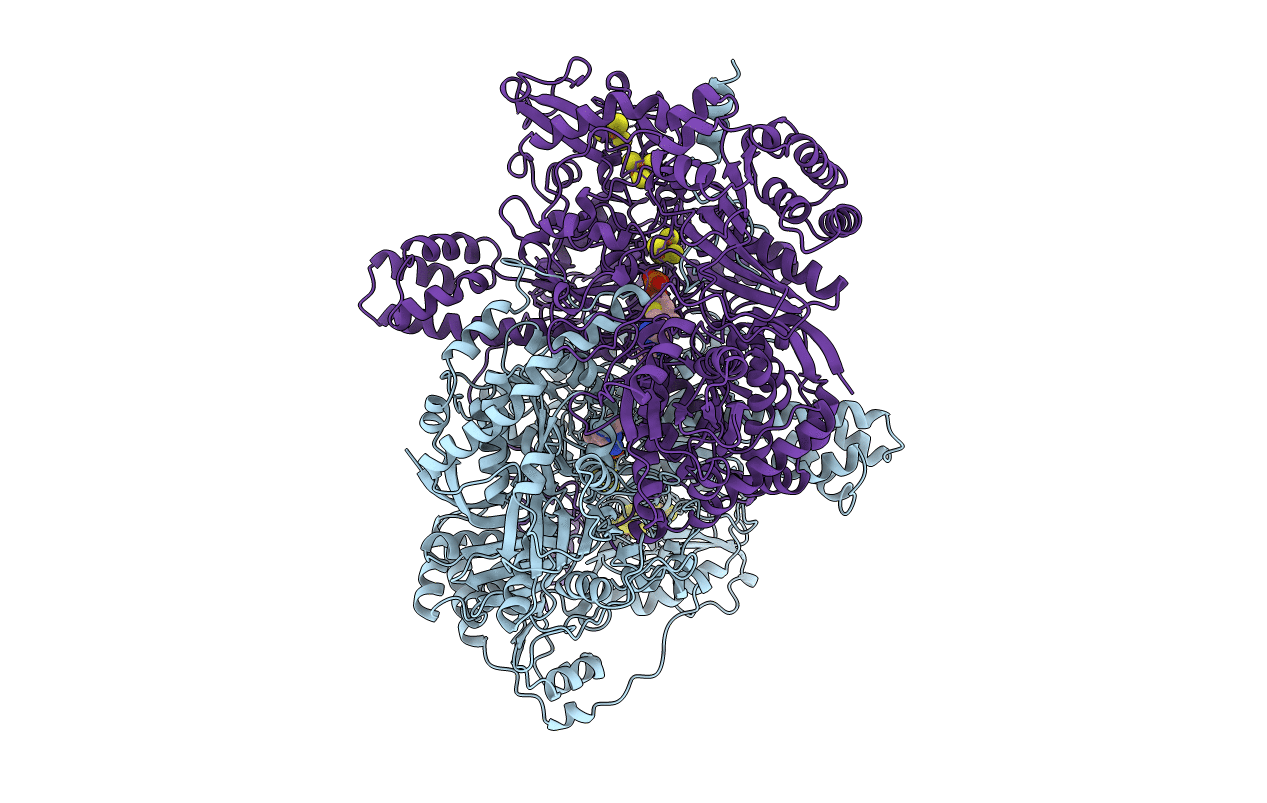
PDB ID:
1B0P
Keywords:
Title:
CRYSTAL STRUCTURE OF PYRUVATE-FERREDOXIN OXIDOREDUCTASE FROM DESULFOVIBRIO AFRICANUS
Biological Source:
Source Organism(s):
Desulfovibrio africanus (Taxon ID: 873)
Method Details:
Experimental Method:
Resolution:
2.31 Å
R-Value Free:
0.27
R-Value Work:
0.19
Space Group:
P 21 21 21
