Deposition Date
1998-11-06
Release Date
2000-02-18
Last Version Date
2024-06-05
Entry Detail
PDB ID:
1B0B
Keywords:
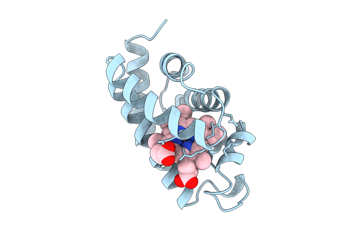
Title:
HEMOGLOBIN I FROM THE CLAM LUCINA PECTINATA, CYANIDE COMPLEX AT 100 KELVIN
Biological Source:
Source Organism(s):
Lucina pectinata (Taxon ID: 29163)
Method Details:
Experimental Method:
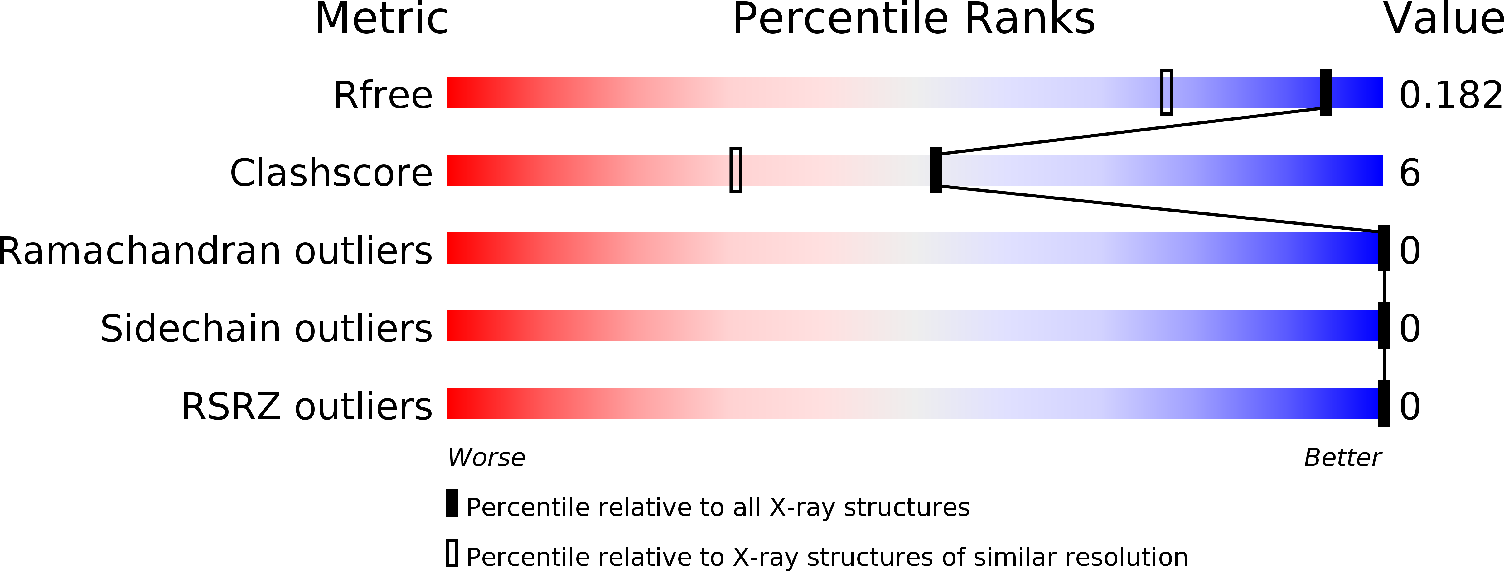
Resolution:
1.43 Å
R-Value Free:
0.16
R-Value Observed:
0.11
Space Group:
P 1 21 1