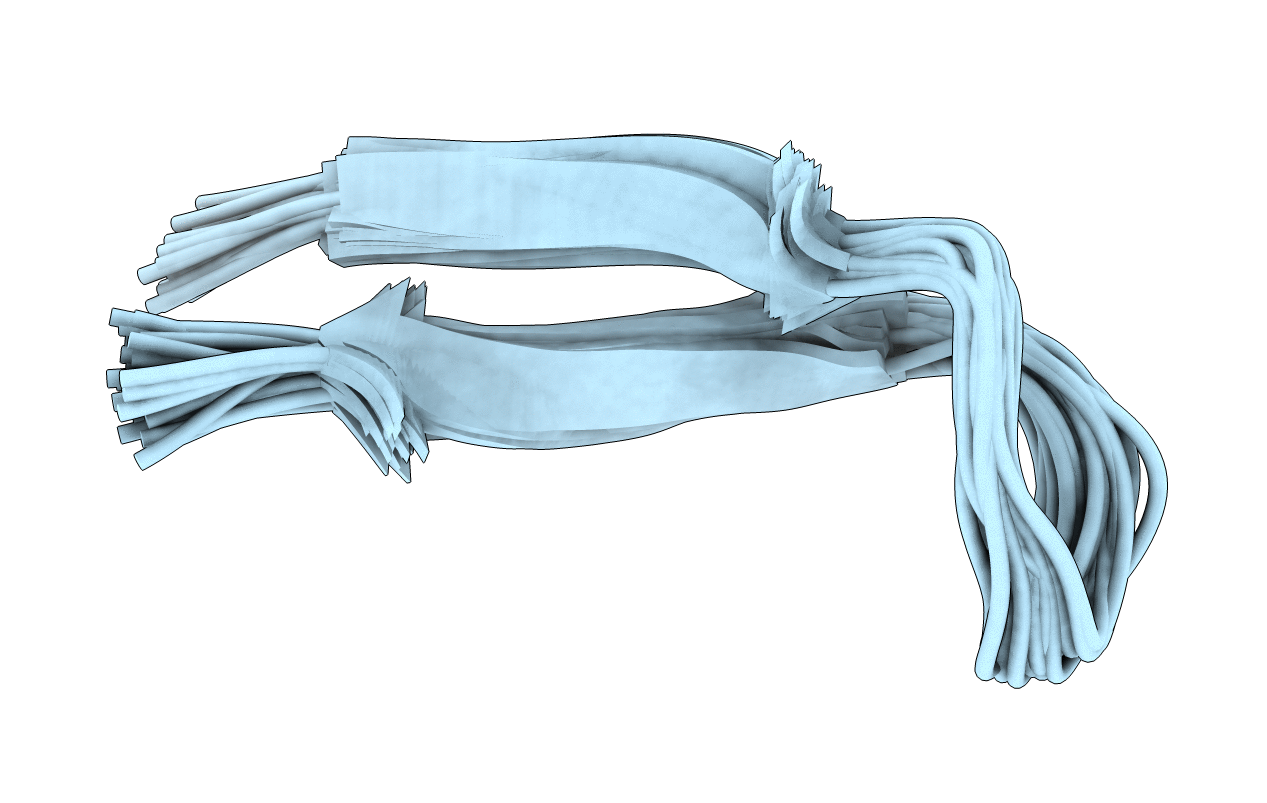
1B03 image
Deposition Date 1998-11-17
Release Date 1998-11-25
Last Version Date 2023-12-27
Entry Detail
PDB ID:
1B03
Keywords:
Title:
SOLUTION STRUCTURE OF THE ANTIBODY-BOUND HIV-1IIIB V3 PEPTIDE
Biological Source:
Source Organism:
Method Details:
Experimental Method:
Conformers Submitted:
35
Selection Criteria:
LEAST RESTRAINT VIOLATIONS
Macromolecular Entities
Structures with similar UniProt ID
Protein Blast
Polymer Type:polypeptide(L)
Molecule:PROTEIN (P1053 PEPTIDE)
Gene (Uniprot):env
Chain IDs:A
Chain Length:18
Number of Molecules:1
Biological Source:
Ligand Molecules
Primary Citation
A cis proline turn linking two beta-hairpin strands in the solution structure of an antibody-bound HIV-1IIIB V3 peptide.
Nat.Struct.Biol. 6 331 335 (1999)
PMID: 10201400 DOI: 10.1038/7567

Abstact

The refined solution structure of an 18-residue HIV-1IIIB V3 peptide in complex with the Fv fragment of an anti-gp120 antibody reveals an unexpected type VI beta-turn comprising residues RGPG at the center of a beta-hairpin. The central glycine and proline of this turn are linked by a cis peptide bond. The residues of the turn interact extensively with the antibody Fv. 15N[1H] NOE measurements show that the backbone of the peptide, including the central QRGPGR loop, is well ordered in the complex. The solution structure is significantly different from the X-ray structures of HIV-1MN V3 peptides bound to anti-peptide antibodies. These differences could be due to a two-residue (QR) insertion preceding the GPGR sequence in the HIV-1IIIB strain, and the much longer peptide epitope immobilized by the anti-gp120 antibody.

Legend

Protein

Chemical

Disease

Primary Citation of related structures