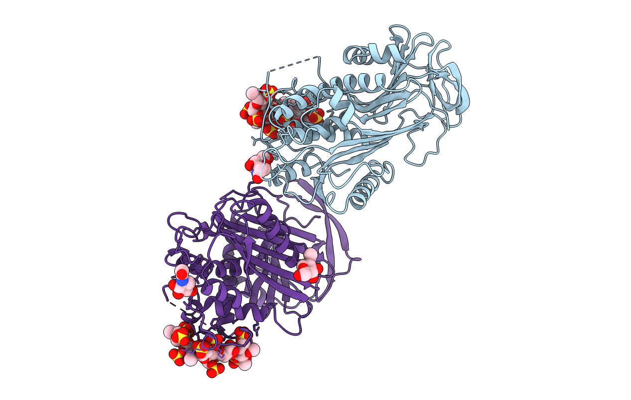
1AZX image
Deposition Date 1997-11-23
Release Date 1999-01-13
Last Version Date 2024-10-30
Entry Detail
PDB ID:
1AZX
Title:
ANTITHROMBIN/PENTASACCHARIDE COMPLEX
Biological Source:
Source Organism:
Homo sapiens (Taxon ID: 9606)
Method Details:
Experimental Method:
Resolution:
2.90 Å
R-Value Free:
0.28
R-Value Work:
0.19
R-Value Observed:
0.20
Space Group:
P 1 21 1
Macromolecular Entities
Polymer Type:polypeptide(L)
Molecule:ANTITHROMBIN
Gene (Uniprot):SERPINC1
Chain IDs:A (auth: I), B (auth: L)
Chain Length:432
Number of Molecules:2
Biological Source:Homo sapiens
Modified Residue
Compound ID Chain ID Parent Comp ID Details 2D Image
ASN A ASN GLYCOSYLATION SITE
Ligand Molecules
Peptide-like Molecules
PRD_900031
Primary Citation
The anticoagulant activation of antithrombin by heparin.
Proc.Natl.Acad.Sci.USA 94 14683 14688 (1997)
PMID: 9405673 DOI: 10.1073/pnas.94.26.14683

Abstact

Antithrombin, a plasma serpin, is relatively inactive as an inhibitor of the coagulation proteases until it binds to the heparan side chains that line the microvasculature. The binding specifically occurs to a core pentasaccharide present both in the heparans and in their therapeutic derivative heparin. The accompanying conformational change of antithrombin is revealed in a 2.9-A structure of a dimer of latent and active antithrombins, each in complex with the high-affinity pentasaccharide. Inhibitory activation results from a shift in the main sheet of the molecule from a partially six-stranded to a five-stranded form, with extrusion of the reactive center loop to give a more exposed orientation. There is a tilting and elongation of helix D with the formation of a 2-turn helix P between the C and D helices. Concomitant conformational changes at the heparin binding site explain both the initial tight binding of antithrombin to the heparans and the subsequent release of the antithrombin-protease complex into the circulation. The pentasaccharide binds by hydrogen bonding of its sulfates and carboxylates to Arg-129 and Lys-125 in the D-helix, to Arg-46 and Arg-47 in the A-helix, to Lys-114 and Glu-113 in the P-helix, and to Lys-11 and Arg-13 in a cleft formed by the amino terminus. This clear definition of the binding site will provide a structural basis for developing heparin analogues that are more specific toward their intended target antithrombin and therefore less likely to exhibit side effects.

Legend

Protein

Chemical

Disease

Primary Citation of related structures