1AZS image
Deposition Date 1997-11-20
Release Date 1998-02-25
Last Version Date 2024-05-22
Entry Detail
PDB ID:
1AZS
Title:
COMPLEX OF GS-ALPHA WITH THE CATALYTIC DOMAINS OF MAMMALIAN ADENYLYL CYCLASE
Biological Source:
Source Organism:
Method Details:
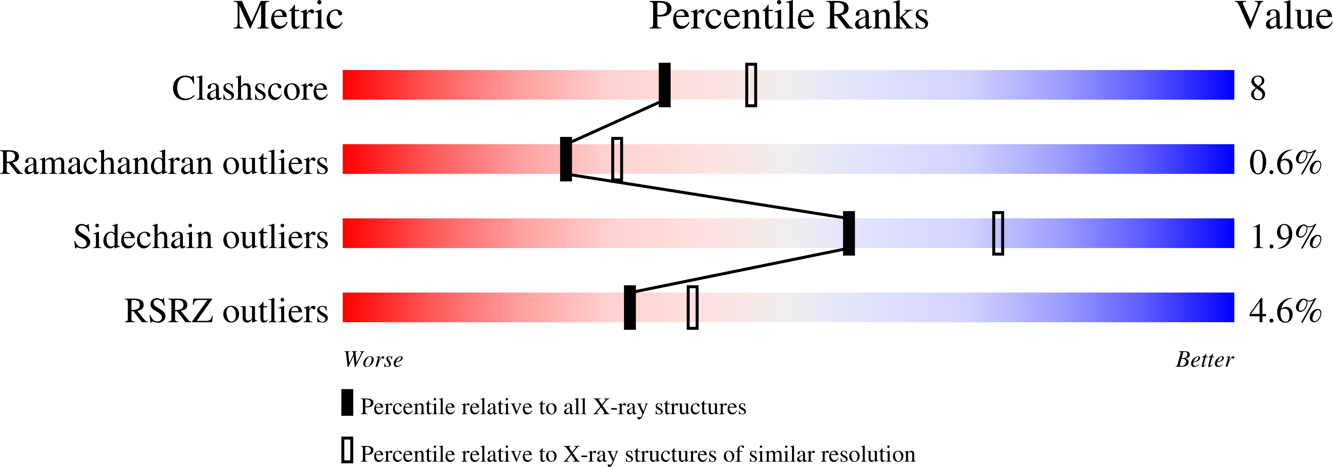
Experimental Method:
Resolution:
2.30 Å
R-Value Free:
0.28
R-Value Work:
0.21
R-Value Observed:
0.21
Space Group:
P 21 21 2
Macromolecular Entities
Structures with similar UniProt ID
Protein Blast
Polymer Type:polypeptide(L)
Molecule:VC1
Gene (Uniprot):ADCY5
Mutagens:V476M, N-TERMINAL HEXAHISTIDINE TAG
Chain IDs:A
Chain Length:220
Number of Molecules:1
Biological Source:Canis lupus familiaris
Structures with similar UniProt ID
Protein Blast
Polymer Type:polypeptide(L)
Molecule:IIC2
Gene (Uniprot):Adcy2
Chain IDs:B
Chain Length:212
Number of Molecules:1
Biological Source:Rattus norvegicus
Structures with similar UniProt ID
Protein Blast
Polymer Type:polypeptide(L)
Molecule:GS-ALPHA
Gene (Uniprot):GNAS
Mutagens:C-TERMINAL HEXAHISTIDINE TAG, NOT PALMITOYLATED AT AMINO TERMINUS
Chain IDs:C
Chain Length:402
Number of Molecules:1
Biological Source:Bos taurus
Primary Citation
Crystal structure of the catalytic domains of adenylyl cyclase in a complex with Gsalpha.GTPgammaS.
Science 278 1907 1916 (1997)
PMID: 9417641 DOI: 10.1126/science.278.5345.1907

Abstact

The crystal structure of a soluble, catalytically active form of adenylyl cyclase in a complex with its stimulatory heterotrimeric G protein alpha subunit (Gsalpha) and forskolin was determined to a resolution of 2.3 angstroms. When P-site inhibitors were soaked into native crystals of the complex, the active site of adenylyl cyclase was located and structural elements important for substrate recognition and catalysis were identified. On the basis of these and other structures, a molecular mechanism is proposed for the activation of adenylyl cyclase by Gsalpha.

Legend

Protein

Chemical

Disease

Primary Citation of related structures
Feedback Form
Name
Email
Institute
Feedback