Deposition Date
1997-11-25
Release Date
1998-05-27
Last Version Date
2024-04-03
Entry Detail
PDB ID:
1AZ5
Keywords:
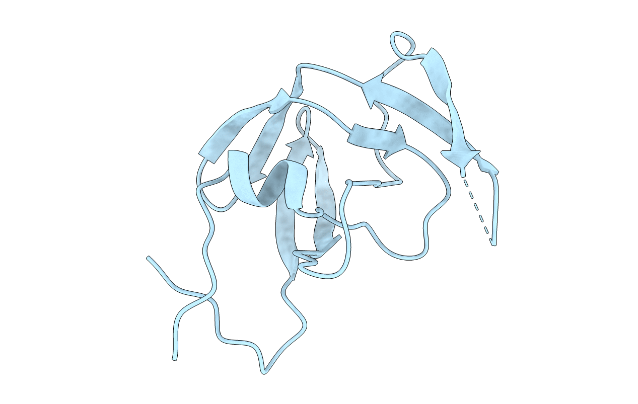
Title:
UNLIGANDED SIV PROTEASE STRUCTURE IN AN "OPEN" CONFORMATION
Biological Source:
Source Organism(s):
Simian immunodeficiency virus (Taxon ID: 11723)
Expression System(s):
Method Details:
Experimental Method:
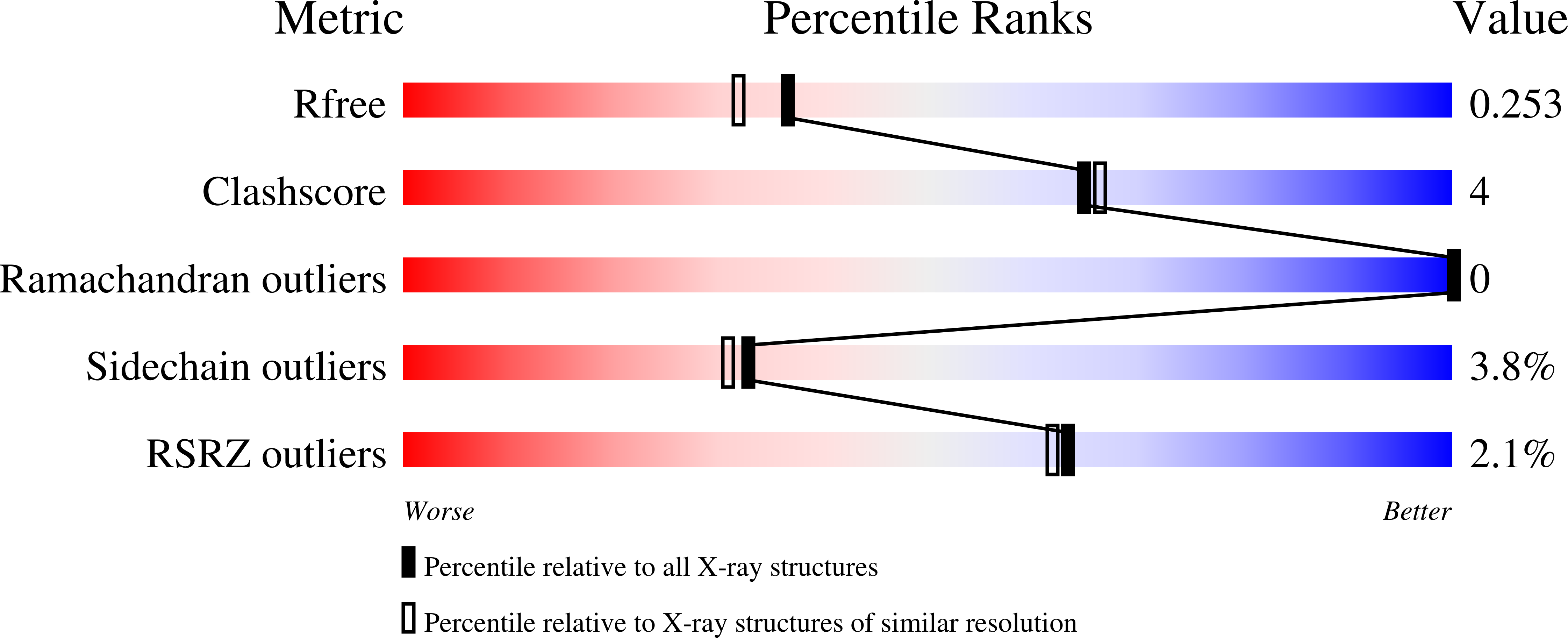
Resolution:
2.00 Å
R-Value Free:
0.26
R-Value Work:
0.20
R-Value Observed:
0.20
Space Group:
P 32 2 1