Deposition Date
1995-08-21
Release Date
1996-03-08
Last Version Date
2024-02-07
Entry Detail
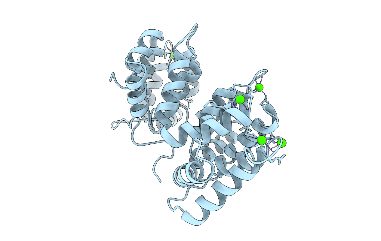
PDB ID:
1AXN
Keywords:
Title:
THE HIGH RESOLUTION STRUCTURE OF ANNEXIN III SHOWS DIFFERENCES WITH ANNEXIN V
Biological Source:
Source Organism(s):
Homo sapiens (Taxon ID: 9606)
Expression System(s):
Method Details:
Experimental Method:
Resolution:
1.78 Å
R-Value Free:
0.22
R-Value Observed:
0.17
Space Group:
P 1 21 1