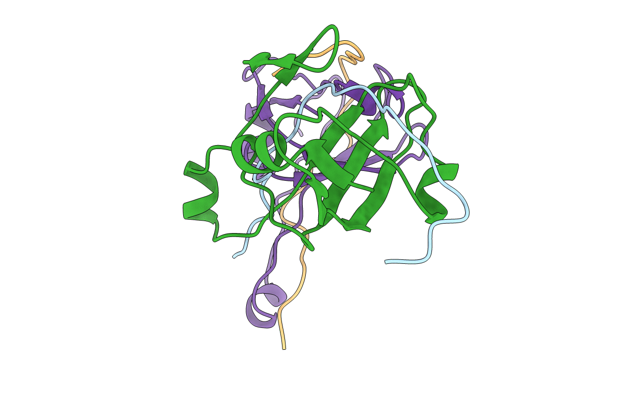
1AW8 image
Entry Detail
PDB ID:
1AW8
Keywords:
Title:
PYRUVOYL DEPENDENT ASPARTATE DECARBOXYLASE
Biological Source:
Source Organism:
Host Organism:
PDB Version:
Deposition Date:
1997-10-12
Release Date:
1998-04-29
Method Details:
Experimental Method:
Resolution:
2.20 Å
R-Value Free:
0.23
R-Value Work:
0.2
R-Value Observed:
0.2
Space Group:
P 61 2 2
Macromolecular Entities
Polymer Type:polypeptide(L)
Description:L-ASPARTATE-ALPHA-DECARBOXYLASE
Chain IDs:A, C (auth: D)
Chain Length:24
Number of Molecules:2
Biological Source:Escherichia coli
Polymer Type:polypeptide(L)
Description:L-ASPARTATE-ALPHA-DECARBOXYLASE
Chain IDs:B, D (auth: E)
Chain Length:91
Number of Molecules:2
Biological Source:Escherichia coli
Primary Citation
Crystal structure of aspartate decarboxylase at 2.2 A resolution provides evidence for an ester in protein self-processing.
Nat.Struct.Biol. 5 289 293 (1998)
PMID: 9546220 DOI: 10.1038/nsb0498-289

Abstact

The structure of L-aspartate-alpha-decarboxylase from E. coli has been determined at 2.2 A resolution. The enzyme is a tetramer with pseudofour-fold rotational symmetry. The subunits are six-stranded beta-barrels capped by small alpha-helices at each end. The active sites are located between adjacent subunits. The electron density provides evidence for catalytic pyruvoyl groups at three active sites and an ester at the fourth. The ester is an intermediate in the autocatalytic self-processing leading to formation of the pyruvoyl group. This unprecedented structure provides novel insights into the general phenomenon of protein processing.

Legend

Protein

Chemical

Disease

Primary Citation of related structures