1AVP image
Deposition Date 1996-04-04
Release Date 1997-11-19
Last Version Date 2024-11-20
Entry Detail
PDB ID:
1AVP
Keywords:
Title:
STRUCTURE OF HUMAN ADENOVIRUS 2 PROTEINASE WITH ITS 11 AMINO ACID COFACTOR
Biological Source:
Source Organism:
Method Details:
Experimental Method:
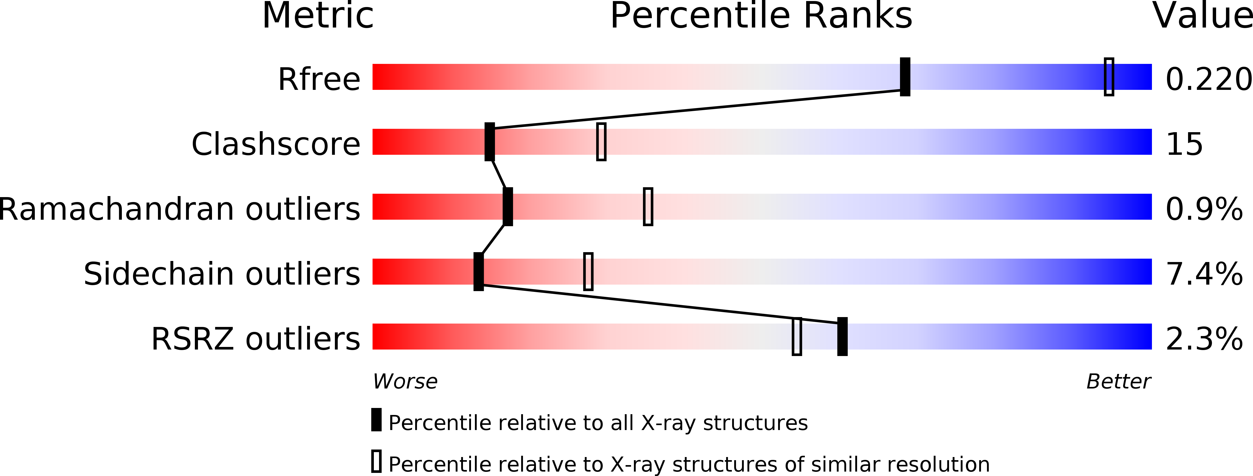
Resolution:
2.60 Å
R-Value Free:
0.23
R-Value Work:
0.18
R-Value Observed:
0.18
Space Group:
P 61
Macromolecular Entities
Polymer Type:polypeptide(L)
Molecule:ADENOVIRAL PROTEINASE
Gene (Uniprot):L3
Chain IDs:A
Chain Length:204
Number of Molecules:1
Biological Source:Human adenovirus 2
Polymer Type:polypeptide(L)
Molecule:ADENOVIRAL PROTEINASE
Gene (Uniprot):L3
Chain IDs:B
Chain Length:11
Number of Molecules:1
Biological Source:Human adenovirus C
Primary Citation
Crystal structure of the human adenovirus proteinase with its 11 amino acid cofactor.
EMBO J. 15 1778 1783 (1996)
PMID: 8617222

Abstact

The three-dimensional structure of the human adenovirus-2 proteinase complexed with its 11 amino acid cofactor, pVIc, was determined at 2.6 A resolution by X-ray crystallographic analysis. The fold of this protein has not been seen before. However, it represents an example of either subtly divergent or powerfully convergent evolution, because the active site contains a Cys-His-Glu triplet and oxyanion hole in an arrangement similar to that in papain. Thus, the adenovirus proteinase represents a new, fifth group of enzymes that contain catalytic triads. pVIc, which extends a beta-sheet in the main chain, is distant from the active site, yet its binding increases the catalytic rate constant 300-fold for substrate hydrolysis. The structure reveals several potential targets for antiviral therapy.

Legend

Protein

Chemical

Disease

Primary Citation of related structures