Deposition Date
1993-03-05
Release Date
1994-01-31
Last Version Date
2024-10-16
Entry Detail
PDB ID:
1AVD
Keywords:
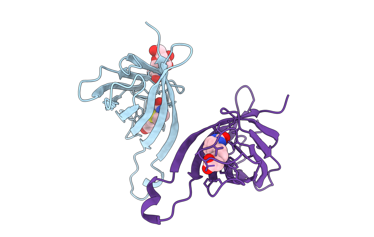
Title:
THREE-DIMENSIONAL STRUCTURE OF THE TETRAGONAL CRYSTAL FORM OF EGG-WHITE AVIDIN IN ITS FUNCTIONAL COMPLEX WITH BIOTIN AT 2.7 ANGSTROMS RESOLUTION
Biological Source:
Source Organism(s):
Gallus gallus (Taxon ID: 9031)
Method Details:
Experimental Method:
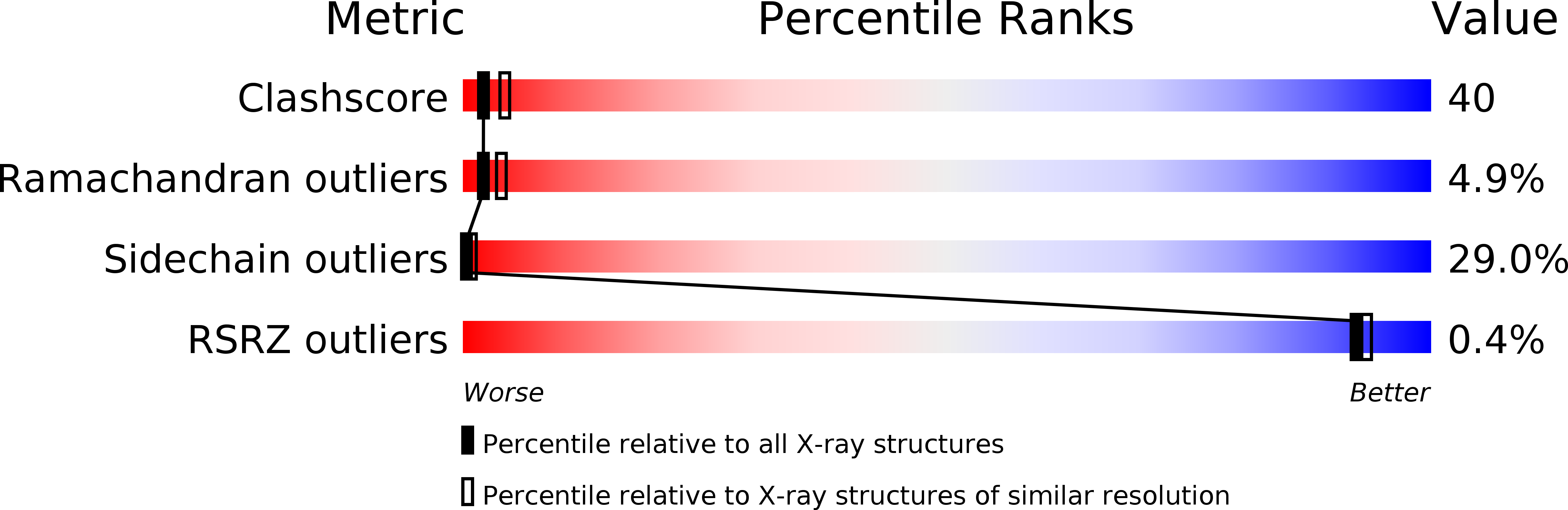
Resolution:
2.70 Å
R-Value Observed:
0.17
Space Group:
P 42 21 2