1AUX image
Deposition Date 1997-09-06
Release Date 1998-03-18
Last Version Date 2024-05-22
Entry Detail
PDB ID:
1AUX
Keywords:
Title:
STRUCTURE OF THE C DOMAIN OF SYNAPSIN IA FROM BOVINE BRAIN WITH CALCIUM ATP-GAMMA-S BOUND
Biological Source:
Source Organism:
Bos taurus (Taxon ID: 9913)
Host Organism:
Method Details:
Experimental Method:
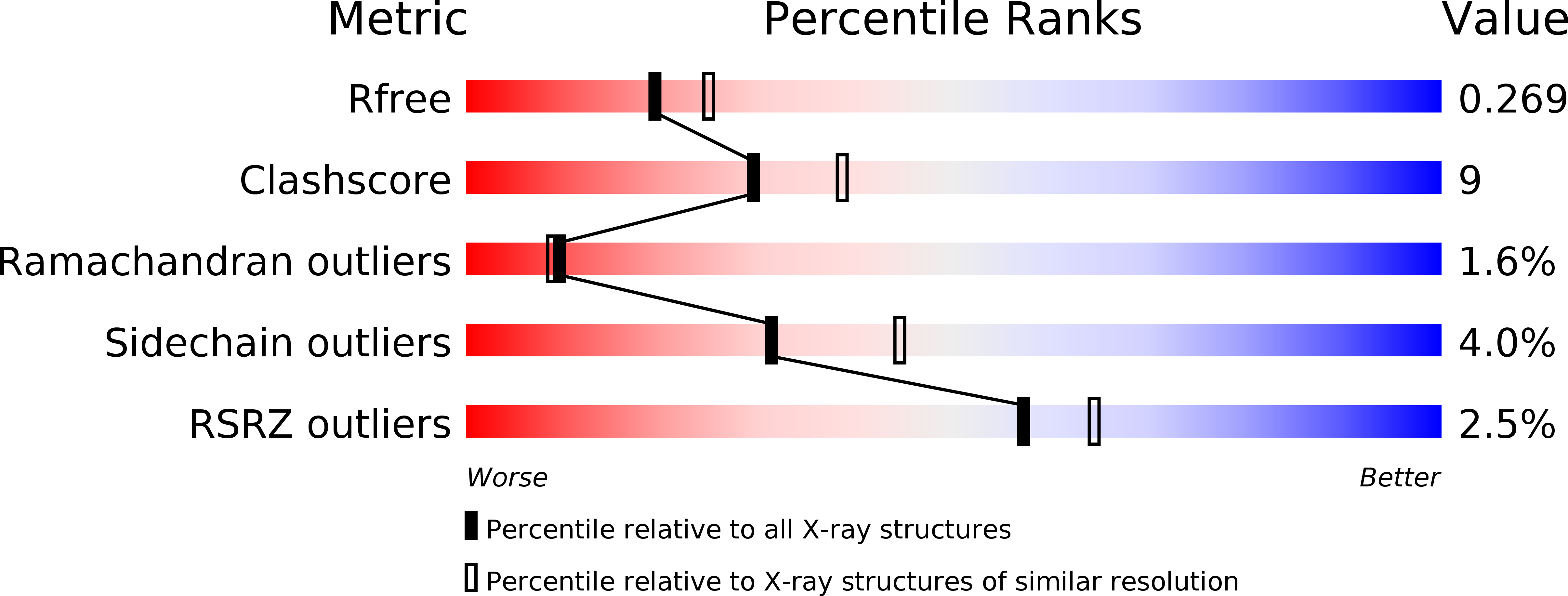
Resolution:
2.30 Å
R-Value Free:
0.26
R-Value Work:
0.20
R-Value Observed:
0.20
Space Group:
P 32 2 1
Macromolecular Entities
Polymer Type:polypeptide(L)
Molecule:SYNAPSIN IA
Gene (Uniprot):SYN1
Chain IDs:A, B
Chain Length:311
Number of Molecules:2
Biological Source:Bos taurus
Primary Citation

Abstact

Synapsins are abundant synaptic vesicle proteins with an essential regulatory function in the nerve terminal. We determined the crystal structure of a fragment (synC) consisting of residues 110-420 of bovine synapsin I; synC coincides with the large middle domain (C-domain), the most conserved domain of synapsins. SynC molecules are folded into compact domains and form closely associated dimers. SynC monomers are strikingly similar in structure to a family of ATP-utilizing enzymes, which includes glutathione synthetase and D-alanine:D-alanine ligase. SynC binds ATP in a Ca2+-dependent manner. The crystal structure of synC in complex with ATPgammaS and Ca2+ explains the preference of synC for Ca2+ over Mg2+. Our results suggest that synapsins may also be ATP-utilizing enzymes.

Legend

Protein

Chemical

Disease

Primary Citation of related structures