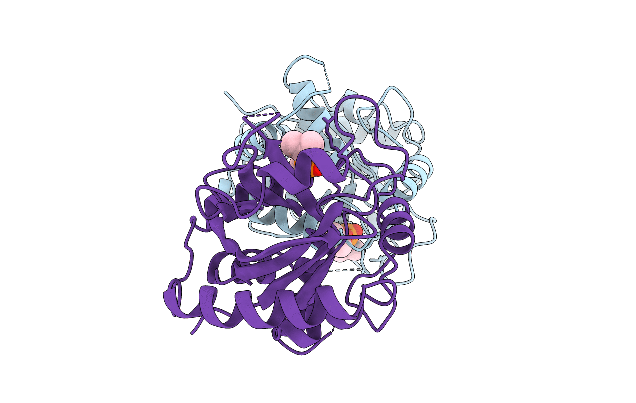
1AT3 image
Deposition Date 1997-08-16
Release Date 1998-10-14
Last Version Date 2024-10-16
Entry Detail
PDB ID:
1AT3
Keywords:
Title:
HERPES SIMPLEX VIRUS TYPE II PROTEASE
Biological Source:
Source Organism:
Host Organism:
Method Details:
Experimental Method:
Resolution:
2.50 Å
R-Value Free:
0.29
R-Value Work:
0.20
R-Value Observed:
0.20
Space Group:
P 21 21 2
Macromolecular Entities
Polymer Type:polypeptide(L)
Molecule:HERPES SIMPLEX VIRUS TYPE II PROTEASE
Gene (Uniprot):UL26
Chain IDs:A, B
Chain Length:247
Number of Molecules:2
Biological Source:Human herpesvirus 2
Ligand Molecules
Primary Citation
Active site cavity of herpesvirus proteases revealed by the crystal structure of herpes simplex virus protease/inhibitor complex.
Biochemistry 36 14023 14029 (1997)
PMID: 9369473 DOI: 10.1021/bi9712697

Abstact

Human herpes simplex virus type 1 (HSV-1) and type 2 (HSV-2) are responsible for herpes labialis (cold sores) and genital herpes, respectively. They encode a serine protease that is required for viral replication, and represent a viable target for therapeutic intervention. Here, we report the crystal structures of HSV-1 and HSV-2 proteases, the latter in the presence and absence of the covalently bound transition state analog inhibitor diisopropyl phosphate (DIP). The HSV-1 and HSV-2 protease structures show a fold that is neither like chymotrypsin nor like subtilisin, and has been seen only in the recently determined cytomegalovirus (CMV) and varicella-zoster virus (VZV) protease structures. HSV-1 and HSV-2 proteases share high sequence homology and have almost identical three-dimensional structures. However, structural differences are observed with the less homologous CMV protease, offering a structural basis for herpes virus protease ligand specificity. The bound inhibitor identifies the oxyanion hole of these enzymes and defines the active site cavity.

Legend

Protein

Chemical

Disease

Primary Citation of related structures