1AST image
Entry Detail
PDB ID:
1AST
Title:
STRUCTURE OF ASTACIN AND IMPLICATIONS FOR ACTIVATION OF ASTACINS AND ZINC-LIGATION OF COLLAGENASES
Biological Source:
Source Organism:
PDB Version:
Deposition Date:
1993-04-21
Release Date:
1994-07-31
Method Details:
Experimental Method:
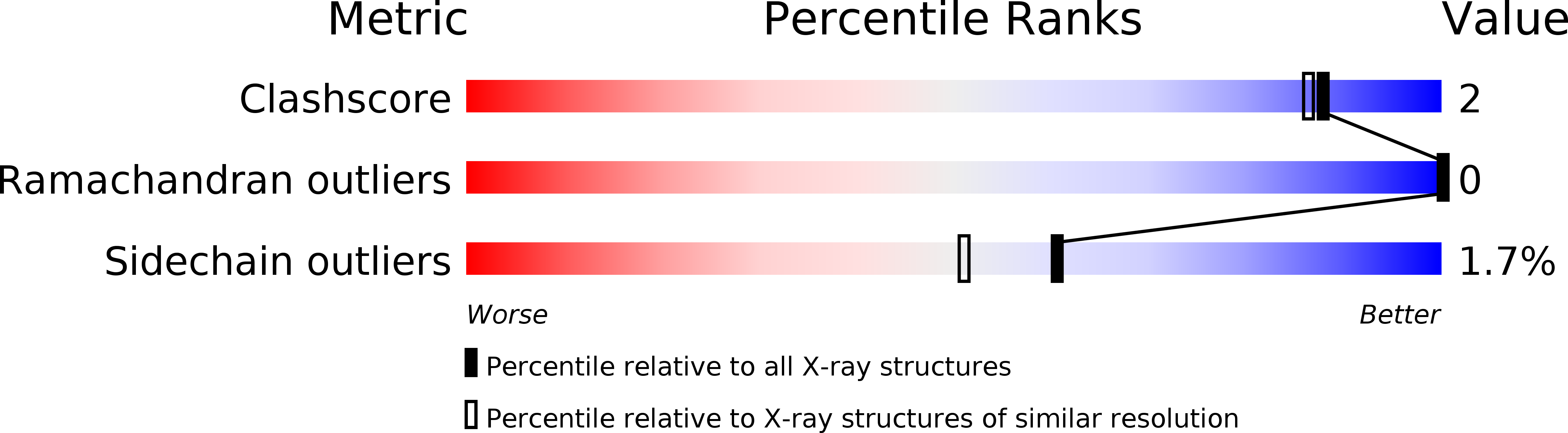
Resolution:
1.80 Å
R-Value Work:
0.15
R-Value Observed:
0.15
Space Group:
P 31 2 1
Macromolecular Entities
Polymer Type:polypeptide(L)
Description:ASTACIN
Chain IDs:A
Chain Length:200
Number of Molecules:1
Biological Source:Astacus astacus
Ligand Molecules
Primary Citation
Structure of astacin and implications for activation of astacins and zinc-ligation of collagenases.
Nature 358 164 167 (1992)
PMID: 1319561 DOI: 10.1038/358164a0

Abstact

Astacin, a digestive zinc-endopeptidase from the crayfish Astacus astacus L., is the prototype for the 'astacin family', which includes mammalian metallo-endopeptidases and developmentally regulated proteins of man, fruitfly, frog and sea urchin. Here we report the X-ray crystal structure of astacin, which reveals a deep active-site cleft, with the zinc at its bottom ligated by three histidines, a water molecule and a more remote tyrosine. The third histidine (His 102) forms part of a consensus sequence, shared not only by the members of the astacin family, but also by otherwise sequentially unrelated proteinases, such as vertebrate collagenases. It may therefore represent the elusive 'third' zinc ligand in these enzymes. The amino terminus of astacin is buried forming an internal salt-bridge with Glu 103, adjacent to His 102. Astacin pro-forms extended at the N terminus, as observed for some 'latent' mammalian astacin homologues, did not exhibit this 'active' conformation, indicating an activation mechanism reminiscent of trypsin-like serine proteinases.

Legend

Protein

Chemical

Disease

Primary Citation of related structures