
Deposition Date
1997-08-08
Release Date
1998-10-14
Last Version Date
2024-05-22
Entry Detail
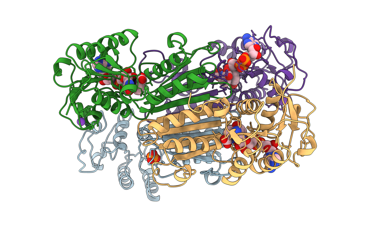
PDB ID:
1ARZ
Keywords:
Title:
ESCHERICHIA COLI DIHYDRODIPICOLINATE REDUCTASE IN COMPLEX WITH NADH AND 2,6 PYRIDINE DICARBOXYLATE
Biological Source:
Source Organism(s):
Escherichia coli (Taxon ID: 83333)
Expression System(s):
Method Details:
Experimental Method:
Resolution:
2.60 Å
R-Value Free:
0.29
R-Value Work:
0.21
R-Value Observed:
0.21
Space Group:
P 21 21 21
