Deposition Date
1993-06-18
Release Date
1994-01-31
Last Version Date
2024-10-30
Entry Detail
PDB ID:
1ARP
Keywords:
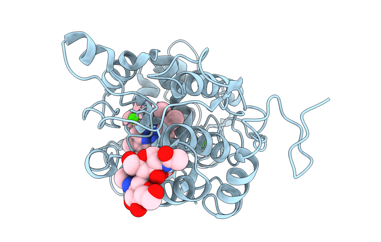
Title:
Crystal structure of the fungal peroxidase from Arthromyces ramosus at 1.9 angstroms resolution: structural comparisons with the lignin and cytochrome C peroxidases
Biological Source:
Source Organism(s):
Penicillium janthinellum (Taxon ID: 5079)
Method Details:
Experimental Method:
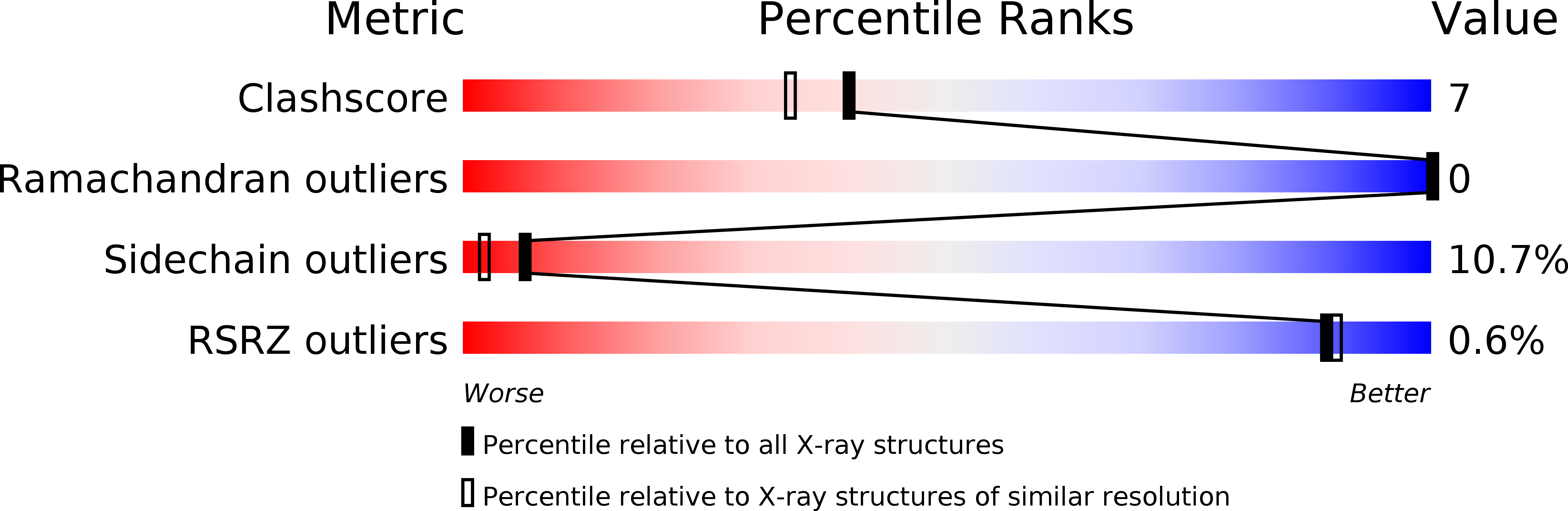
Resolution:
1.90 Å
R-Value Work:
0.17
R-Value Observed:
0.17
Space Group:
P 42 21 2