Deposition Date
1997-08-04
Release Date
1997-11-12
Last Version Date
2024-10-30
Entry Detail
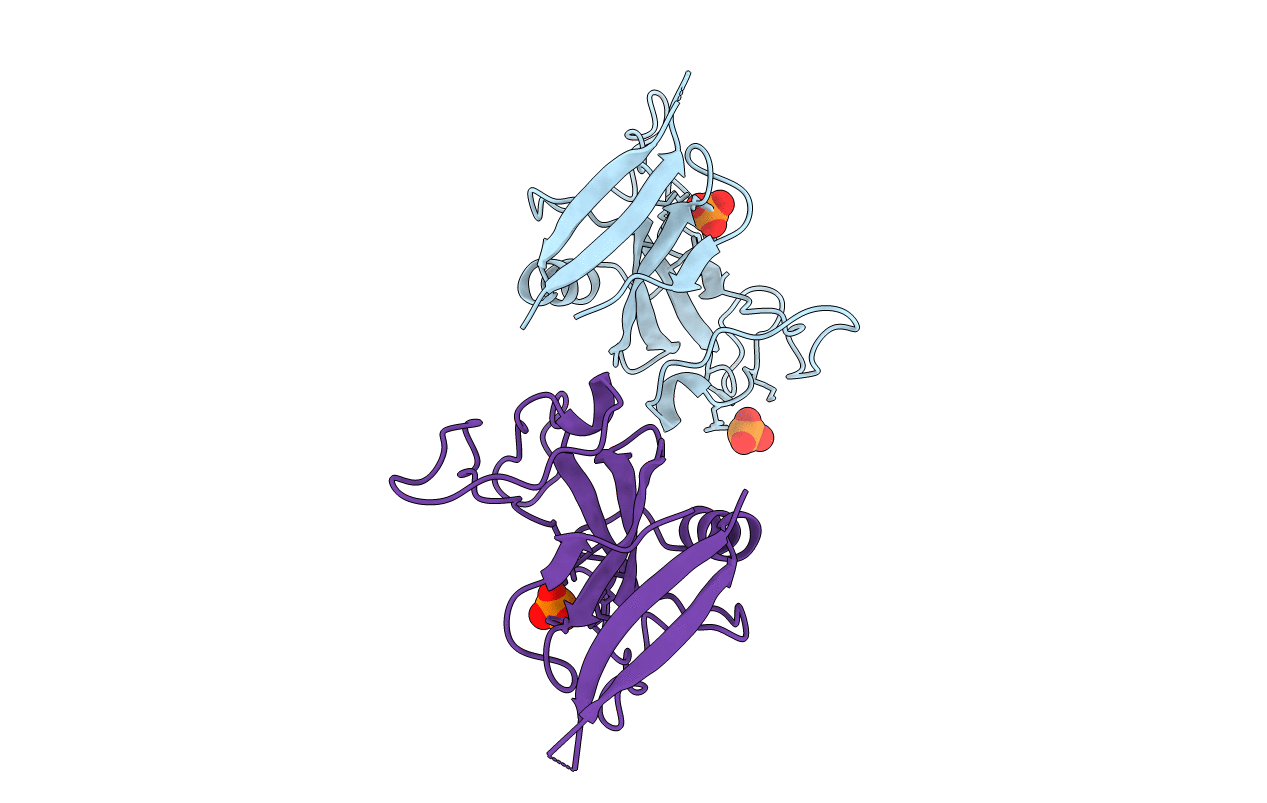
PDB ID:
1AQZ
Keywords:
Title:
CRYSTAL STRUCTURE OF A HIGHLY SPECIFIC ASPERGILLUS RIBOTOXIN, RESTRICTOCIN
Biological Source:
Source Organism(s):
Aspergillus restrictus (Taxon ID: 5064)
Method Details:
Experimental Method:
Resolution:
1.70 Å
R-Value Free:
0.17
R-Value Work:
0.23
R-Value Observed:
0.23
Space Group:
P 1 21 1